File:Lewis1906 fig001.jpg
From Embryology

Size of this preview: 800 × 488 pixels. Other resolution: 1,186 × 724 pixels.
Original file (1,186 × 724 pixels, file size: 132 KB, MIME type: image/jpeg)
Fig. 1. Diagram of a Cell
Microsomes and filar mass only partly sketched.
| Historic Disclaimer - information about historic embryology pages |
|---|
 |
- Links: Stoehr's Histology 1906 | Figures | Historic Embryology | Histology
Reference
Lewis, FT. Stoehr's Histology. (1906) P. Blakiston's Son & Co., Philadelphia.
Cite this page: Hill, M.A. (2024, April 26) Embryology Lewis1906 fig001.jpg. Retrieved from https://embryology.med.unsw.edu.au/embryology/index.php/File:Lewis1906_fig001.jpg
- © Dr Mark Hill 2024, UNSW Embryology ISBN: 978 0 7334 2609 4 - UNSW CRICOS Provider Code No. 00098G
File history
Click on a date/time to view the file as it appeared at that time.
| Date/Time | Thumbnail | Dimensions | User | Comment | |
|---|---|---|---|---|---|
| current | 18:18, 6 February 2015 | | 1,186 × 724 (132 KB) | Z8600021 (talk | contribs) | |
| 18:18, 6 February 2015 | 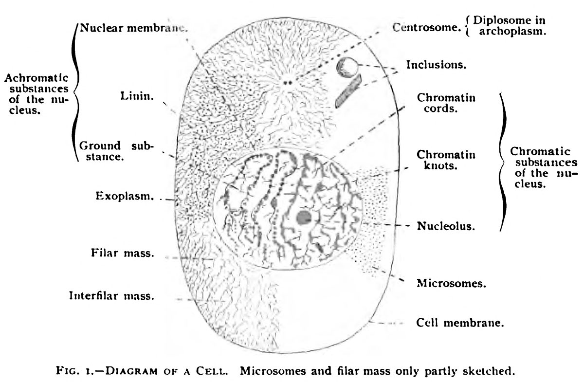 | 1,200 × 785 (144 KB) | Z8600021 (talk | contribs) |
You cannot overwrite this file.
File usage
The following 2 pages use this file: