File:LeeHalpert1932 plate04.jpg

Original file (1,000 × 2,015 pixels, file size: 271 KB, MIME type: image/jpeg)
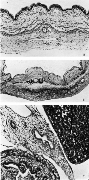
Plate 4 Fetal Gall Bladder
5 Gall bladder of a 370 mm fetus in longitudinal section. The active proliferation of the lining epithelium is rather conspicuous. Photomicrograph, X 120.
6 Gall bladder of a 280 mm fetus in cross section. The coarse ramifications (plexus perimuseularis) of the large vessels are located in the dense zone of the perimuseular layer, in that adjacent to the muscular coat. Branches of these vessels communicate through the muscular layer with the rich capillary bed (plexus subepithelialis) distributed beneath the lining epithelium. Photomier0graph, X 60.
7 The hepatocystic junction of the 40 mm human embryo. The lumen of the cystic duct merges gradually with that of the common hepatic duct. The endodermal portion of both duets is represented by a tube lined with a single row of columnar epithelial cells. The mosodermal portion is wholly undifferentiated. Photomicrograph, X 120.
Reference
Halpert B. and Lee H. The gall bladder and the extrahepatic biliary passages in late embryonic and early fetal life. (1932) Anat. Rec. 54(1): 29-42.
Cite this page: Hill, M.A. (2024, April 26) Embryology LeeHalpert1932 plate04.jpg. Retrieved from https://embryology.med.unsw.edu.au/embryology/index.php/File:LeeHalpert1932_plate04.jpg
- © Dr Mark Hill 2024, UNSW Embryology ISBN: 978 0 7334 2609 4 - UNSW CRICOS Provider Code No. 00098G
File history
Click on a date/time to view the file as it appeared at that time.
| Date/Time | Thumbnail | Dimensions | User | Comment | |
|---|---|---|---|---|---|
| current | 15:26, 29 August 2017 |  | 1,000 × 2,015 (271 KB) | Z8600021 (talk | contribs) | |
| 15:26, 29 August 2017 |  | 1,378 × 2,258 (268 KB) | Z8600021 (talk | contribs) | ===Reference=== {{Ref-LeeHalpert1932}} {{Footer}} Category:Gall BladderCategory:1930's |
You cannot overwrite this file.
File usage
There are no pages that use this file.