File:LeeHalpert1932 plate01.jpg

Original file (1,234 × 2,148 pixels, file size: 247 KB, MIME type: image/jpeg)
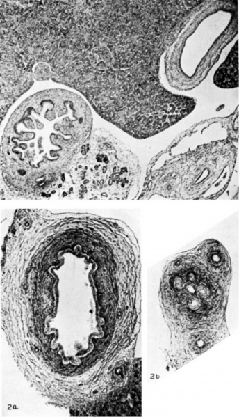
Plate 1 Fetal Gall Bladder
1 Gall bladder and duodenum of a 40 mm human embryo; cross section at the level of the umbilicus. The cells of the endodermal epithelial lining appear uniform in size and staining reaction. The mesodermal portion of the gall bladder shows only a slight indication of differentiation into layers. The passage through the duodenal wall of the ductus choledochus and the ductus pancreaticus is seen. Photomicrograph, X 50.
2 Gall bladder of an 80 mm human embryo; cross sections from the corpus (a.) and from the neck (b). The alveolar indentations and corresponding round elevations give a pattern to the mucosal surface. This is particularly marked in the neck, where the stellate lumen is composed of several communicating epithelial-lined indentations. The muscular coat is a well-defined layer of smooth-muscle cells embedded in a connective-tissue stroma. Photomicrographs, X 60.
- Links: gallbladder
Reference
Halpert B. and Lee H. The gall bladder and the extrahepatic biliary passages in late embryonic and early fetal life. (1932) Anat. Rec. 54(1): 29-42.
Cite this page: Hill, M.A. (2024, April 26) Embryology LeeHalpert1932 plate01.jpg. Retrieved from https://embryology.med.unsw.edu.au/embryology/index.php/File:LeeHalpert1932_plate01.jpg
- © Dr Mark Hill 2024, UNSW Embryology ISBN: 978 0 7334 2609 4 - UNSW CRICOS Provider Code No. 00098G
File history
Click on a date/time to view the file as it appeared at that time.
| Date/Time | Thumbnail | Dimensions | User | Comment | |
|---|---|---|---|---|---|
| current | 15:27, 29 August 2017 |  | 1,234 × 2,148 (247 KB) | Z8600021 (talk | contribs) | |
| 15:26, 29 August 2017 |  | 1,354 × 2,454 (237 KB) | Z8600021 (talk | contribs) | ===Reference=== {{Ref-LeeHalpert1932}} {{Footer}} Category:Gall BladderCategory:1930's |
You cannot overwrite this file.
File usage
There are no pages that use this file.