File:Kollmann758.jpg
Kollmann758.jpg (780 × 406 pixels, file size: 45 KB, MIME type: image/jpeg)
- This text is a Google translate computer generated translation and may contain many errors.
Images from - Atlas of the Development of Man (Volume 2)
(Handatlas der entwicklungsgeschichte des menschen)
- Kollmann Atlas 2: Gastrointestinal | Respiratory | Urogenital | Cardiovascular | Neural | Integumentary | Smell | Vision | Hearing | Kollmann Atlas 1 | Kollmann Atlas 2 | Julius Kollmann
- Links: Julius Kollman | Atlas Vol.1 | Atlas Vol.2 | Embryology History
| Historic Disclaimer - information about historic embryology pages |
|---|
 |
Reference
Kollmann JKE. Atlas of the Development of Man (Handatlas der entwicklungsgeschichte des menschen). (1907) Vol.1 and Vol. 2. Jena, Gustav Fischer. (1898).
Cite this page: Hill, M.A. (2024, April 26) Embryology Kollmann758.jpg. Retrieved from https://embryology.med.unsw.edu.au/embryology/index.php/File:Kollmann758.jpg
- © Dr Mark Hill 2024, UNSW Embryology ISBN: 978 0 7334 2609 4 - UNSW CRICOS Provider Code No. 00098G
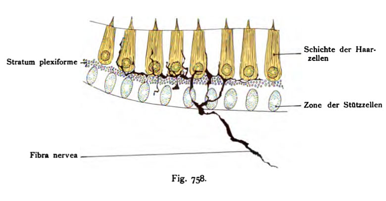
Fig. 758. Nervenfasern in der Schichte der Neuroepithelien der Macula
acustica sacculi
der jungen weißen Maus nach Anwendung Golgis Methode.
(Nach V. Lenhoss^k.)
Ein nackter Achsenzylinder tritt in die Zone der Stützzellen ein, teilt sich, zieht hier in drei Nervenfäden gespalten in die Höhe, welche entweder un- mittelbar zu den gewölbten Enden der Haarzellen laufen oder eine Strecke weit an der Seitenwand der Zellen in die (Höhe steigen. Sie endigen pericellulär mit freien Endspitzen, ohne die freie Oberfläche zu erreichen. Im Stratum plexiforme greifen die feinen Fasern oft innig ineinander.
File history
Click on a date/time to view the file as it appeared at that time.
| Date/Time | Thumbnail | Dimensions | User | Comment | |
|---|---|---|---|---|---|
| current | 12:40, 21 October 2011 | | 780 × 406 (45 KB) | S8600021 (talk | contribs) | {{Kollmann1907}} Category:Hearing Fig. 758. Nervenfasern in der Schichte der Neuroepithelien der Macula acustica sacculi der jungen weißen Maus nach Anwendung Golgis Methode. (Nach V. Lenhoss^k.) Ein nackter Achsenzylinder tritt in die Zo |
You cannot overwrite this file.
File usage
The following page uses this file: