File:Kollmann585.jpg
Kollmann585.jpg (604 × 558 pixels, file size: 39 KB, MIME type: image/jpeg)
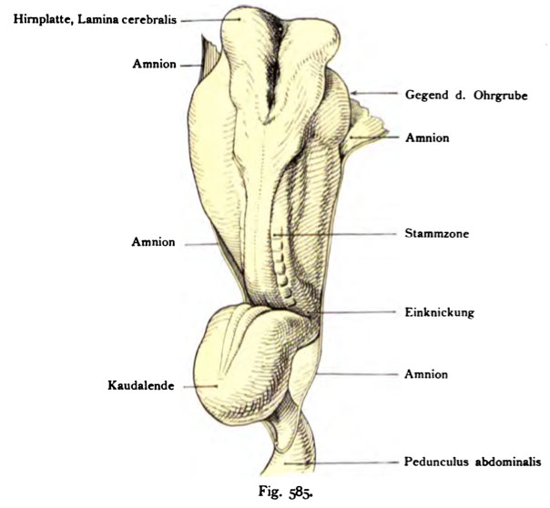
Fig. 585. The formation of the nervous system in a human embryo of 2.69 mm length
measured from the head end to the amnion envelope on the belly stalk.
Dorsal view.
(According to Graf Spee.)
The embryo is still strongly bent. To the piston-like thickened Head is the brain plate, cerebral lamina, dorsal and open in front, bounded by strikingly sculpted medullary In the middle part of the embryo is
the neural tube emerged. On the curved tail of the embryo is the neural canal to a "neural plate, lamina medullaris, set apart wide.
- This text is a Google translate computer generated translation and may contain many errors.
Images from - Atlas of the Development of Man (Volume 2)
(Handatlas der entwicklungsgeschichte des menschen)
- Kollmann Atlas 2: Gastrointestinal | Respiratory | Urogenital | Cardiovascular | Neural | Integumentary | Smell | Vision | Hearing | Kollmann Atlas 1 | Kollmann Atlas 2 | Julius Kollmann
- Links: Julius Kollman | Atlas Vol.1 | Atlas Vol.2 | Embryology History
| Historic Disclaimer - information about historic embryology pages |
|---|
| Pages where the terms "Historic" (textbooks, papers, people, recommendations) appear on this site, and sections within pages where this disclaimer appears, indicate that the content and scientific understanding are specific to the time of publication. This means that while some scientific descriptions are still accurate, the terminology and interpretation of the developmental mechanisms reflect the understanding at the time of original publication and those of the preceding periods, these terms, interpretations and recommendations may not reflect our current scientific understanding. (More? Embryology History | Historic Embryology Papers) |
Reference
Kollmann JKE. Atlas of the Development of Man (Handatlas der entwicklungsgeschichte des menschen). (1907) Vol.1 and Vol. 2. Jena, Gustav Fischer. (1898).
Cite this page: Hill, M.A. (2024, April 26) Embryology Kollmann585.jpg. Retrieved from https://embryology.med.unsw.edu.au/embryology/index.php/File:Kollmann585.jpg
- © Dr Mark Hill 2024, UNSW Embryology ISBN: 978 0 7334 2609 4 - UNSW CRICOS Provider Code No. 00098G
Fig, 585. Die Anläse des Nervensystems bei einem menschlichen Embryo von
2,69 mm Länge,
gemessen vom Kopfende bis zum Amnionsumschlag auf dem Bauchstiel.
Norma dorsalis. (Nach Graf Spee.)
Der Embryo ist noch stark geknickt. An dem kolbenförmig verdickten Kopfteil ist die Hirnplatte, Lamina cerebralis, dorsal und vorn offen, von auffallend modellierten Medullarwülsten begrenzt Im Mittelstück des Embryo ist das MeduUarrohr entstanden. Auf dem gekrümmten Schwanzstück des Embryo ist die MeduUarrinne zu einer „Medullarplatte, Lamina meduUaris, breit ausein- andergelegt.
File history
Click on a date/time to view the file as it appeared at that time.
| Date/Time | Thumbnail | Dimensions | User | Comment | |
|---|---|---|---|---|---|
| current | 16:24, 17 October 2011 |  | 604 × 558 (39 KB) | S8600021 (talk | contribs) | {{Kollmann1907}} |
You cannot overwrite this file.
File usage
The following page uses this file:

