File:Kollmann397.jpg
Original file (862 × 554 pixels, file size: 64 KB, MIME type: image/jpeg)
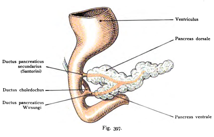
Fig. 397. Pancreas systems in a human embryo by the end of the 6th Week
The glandular systems are now contacted each other and at a location corresponding to that of the mature pancreas, namely in the head region, where the pancreatic duct secundarius (Santorini) of the pancreatic duct (Wirsungi) branches.
- This text is a Google translate computer generated translation and may contain many errors.
Images from - Atlas of the Development of Man (Volume 2)
(Handatlas der entwicklungsgeschichte des menschen)
- Kollmann Atlas 2: Gastrointestinal | Respiratory | Urogenital | Cardiovascular | Neural | Integumentary | Smell | Vision | Hearing | Kollmann Atlas 1 | Kollmann Atlas 2 | Julius Kollmann
- Links: Julius Kollman | Atlas Vol.1 | Atlas Vol.2 | Embryology History
| Historic Disclaimer - information about historic embryology pages |
|---|
 |
Reference
Kollmann JKE. Atlas of the Development of Man (Handatlas der entwicklungsgeschichte des menschen). (1907) Vol.1 and Vol. 2. Jena, Gustav Fischer. (1898).
Cite this page: Hill, M.A. (2024, April 26) Embryology Kollmann397.jpg. Retrieved from https://embryology.med.unsw.edu.au/embryology/index.php/File:Kollmann397.jpg
- © Dr Mark Hill 2024, UNSW Embryology ISBN: 978 0 7334 2609 4 - UNSW CRICOS Provider Code No. 00098G
Fig. 397. Panlcreasanlagen bei einem menschlichen Embryo
vom Ende der 6. Woche.
Die Drüsenanlagen sind jetzt miteinander in Verbindung getreten und zwar an einer Stelle, welche derjenigen des reifen Pankreas entspricht, nämlich im Bereich des Kopfes, dort wo der Ductus pancreaticus secundarius (Santorini) von dem Ductus pancreaticus (Wirsungi) abzweigt
File history
Click on a date/time to view the file as it appeared at that time.
| Date/Time | Thumbnail | Dimensions | User | Comment | |
|---|---|---|---|---|---|
| current | 13:48, 16 October 2011 | | 862 × 554 (64 KB) | S8600021 (talk | contribs) | {{Kollmann1907}} |
You cannot overwrite this file.
File usage
The following 2 pages use this file: