File:Kollmann350.jpg

Original file (767 × 669 pixels, file size: 55 KB, MIME type: image/jpeg)
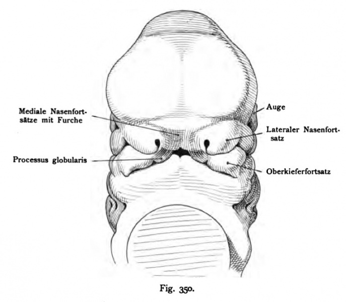
Fig. 350. Face of a human embryo of 11.3 mm (30 - 31 days old) presented en face
(After Rabl)
The head is placed upright as in the adult, because the natural shape of the embryo is the largest part of the face with the heart in touch and so approached, nothing is noticeable that from the face. The comparison with the earlier stages (Fig. 346 and 349) shows that the nasal placodes no longer available as it used the olfactory fields laterally, but are directed straight ahead. The lowermost end of the medial Nasal process, which combines with the maxillary process is, as in rabbit and pig by a shallow impression of the other training abgegliedert rate and represents the process of escalation of the globular head down runs on the medial nasal processes a furrow, which is the mouth-opening on the hard palate continues. The mandibular arch clearly shows the medial division.
- This text is a Google translate computer generated translation and may contain many errors.
Images from - Atlas of the Development of Man (Volume 2)
(Handatlas der entwicklungsgeschichte des menschen)
- Kollmann Atlas 2: Gastrointestinal | Respiratory | Urogenital | Cardiovascular | Neural | Integumentary | Smell | Vision | Hearing | Kollmann Atlas 1 | Kollmann Atlas 2 | Julius Kollmann
- Links: Julius Kollman | Atlas Vol.1 | Atlas Vol.2 | Embryology History
| Historic Disclaimer - information about historic embryology pages |
|---|
| Pages where the terms "Historic" (textbooks, papers, people, recommendations) appear on this site, and sections within pages where this disclaimer appears, indicate that the content and scientific understanding are specific to the time of publication. This means that while some scientific descriptions are still accurate, the terminology and interpretation of the developmental mechanisms reflect the understanding at the time of original publication and those of the preceding periods, these terms, interpretations and recommendations may not reflect our current scientific understanding. (More? Embryology History | Historic Embryology Papers) |
Reference
Kollmann JKE. Atlas of the Development of Man (Handatlas der entwicklungsgeschichte des menschen). (1907) Vol.1 and Vol. 2. Jena, Gustav Fischer. (1898).
Cite this page: Hill, M.A. (2024, April 26) Embryology Kollmann350.jpg. Retrieved from https://embryology.med.unsw.edu.au/embryology/index.php/File:Kollmann350.jpg
- © Dr Mark Hill 2024, UNSW Embryology ISBN: 978 0 7334 2609 4 - UNSW CRICOS Provider Code No. 00098G
Fig. 350. Gesicht eines menschlichen Embryo von 11,3 mm (30 — 31 Tage alt), en face dargestellt.
(Nach RabL)
Der Kopf ist aufrecht gestellt wie bei dem Erwachsenen, denn bei der natürlichen Form des Embryo ist der größte Teil des Antlitzes mit dem Herz- wult in Berührung und so genähert, daß vom Gesicht nichts bemerkbar ist. Die Vergleichung mit den früheren Stufen (Fig. 346 und 349) zeigt, daß die Nasengrübchen nicht mehr, wie früher die Riechfelder lateralwärts stehen, sondern direkt nach vorn gerichtet sind. Das unterste Ende der medialen Nasenfortsätze, welches sich mit dem Oberkieferfortsatz verbindet, ist wie beim Kaninchen und Schwein durch einen seichten Eindruck von dem übrigen Fort- satz abgegliedert und stellt den Processus globularis dar. Von der Stirn herab läuft über die medialen Nasenfortsätze eine Furche, die sich durch die Mund- öffnung auf den harten Gaumen fortsetzt. Der Unterkieferbogen zeigt deutlich die mediale Trennung.
File history
Click on a date/time to view the file as it appeared at that time.
| Date/Time | Thumbnail | Dimensions | User | Comment | |
|---|---|---|---|---|---|
| current | 12:57, 16 October 2011 |  | 767 × 669 (55 KB) | S8600021 (talk | contribs) | {{Kollmann1907}} |
You cannot overwrite this file.
File usage
The following page uses this file:
