File:Kollmann349.jpg

Original file (709 × 657 pixels, file size: 51 KB, MIME type: image/jpeg)
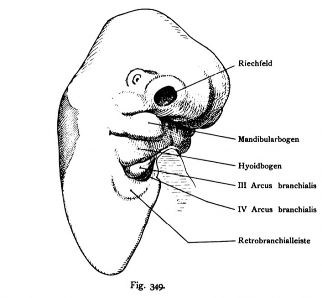
Fig. 349. Head of an Human embryo of 8.3 mm CRL (late 4th or early 5th week) in half-profile
(After Rabl)
The outer shape of the head and especially through the gills and gill-pouches limited foregut, the olfactory fields with the two-Naseii appendages are well developed, partly hidden behind the eye, the through the lens ectoderm is visible through the wide slit-like mouth opening is above the hemisphere surmounted by vesicles. to the site is located to the upper and lower jaw protrusion as parts of Mandibular arch, then follow the rest of the gill arches, the foregut surrounded laterally and ventrally. Third and fourth arches are basically the Sinus located präcervicalis that of a strongly developed brachial bar is moved, as in pigs and rabbits. At the back of the head gleams the fourth ventricle through the skin.
- This text is a Google translate computer generated translation and may contain many errors.
Images from - Atlas of the Development of Man (Volume 2)
(Handatlas der entwicklungsgeschichte des menschen)
- Kollmann Atlas 2: Gastrointestinal | Respiratory | Urogenital | Cardiovascular | Neural | Integumentary | Smell | Vision | Hearing | Kollmann Atlas 1 | Kollmann Atlas 2 | Julius Kollmann
- Links: Julius Kollman | Atlas Vol.1 | Atlas Vol.2 | Embryology History
| Historic Disclaimer - information about historic embryology pages |
|---|
| Pages where the terms "Historic" (textbooks, papers, people, recommendations) appear on this site, and sections within pages where this disclaimer appears, indicate that the content and scientific understanding are specific to the time of publication. This means that while some scientific descriptions are still accurate, the terminology and interpretation of the developmental mechanisms reflect the understanding at the time of original publication and those of the preceding periods, these terms, interpretations and recommendations may not reflect our current scientific understanding. (More? Embryology History | Historic Embryology Papers) |
Reference
Kollmann JKE. Atlas of the Development of Man (Handatlas der entwicklungsgeschichte des menschen). (1907) Vol.1 and Vol. 2. Jena, Gustav Fischer. (1898).
Cite this page: Hill, M.A. (2024, April 26) Embryology Kollmann349.jpg. Retrieved from https://embryology.med.unsw.edu.au/embryology/index.php/File:Kollmann349.jpg
- © Dr Mark Hill 2024, UNSW Embryology ISBN: 978 0 7334 2609 4 - UNSW CRICOS Provider Code No. 00098G
Fig. 349. Kopf eines measchUchen Embryo von 8,3 mm Nackensteißlänge (Ende der 4. oder Anfang der S.Woche) im Halbprofil.
(Nach RabL)
Die äußere Form des Kopfes und besonders des durch Kiemenbogen und
Kiementaschen begrenzten Kopfdarms, die Riechfelder mit den beiden Naseii-
fortsätzen sind gut entwickelt, dahinter das teilweise verdeckte Auge, dessen
Linse durch das Ektoderm hindurch erkennbar ist Die breite spaltförmige
Mundöffnung wird oben her durch die Hemisphärenbläschen überwölbt. An
den Seiten befindet sich der Ober- und der Unterkieferfortsatz als Teile des
Mandibularbogens, dann folgen die übrigen Kiemenbogen, welche den Kopfdarm
seitlich und ventral umgeben. Dritter und vierter Bogen sind im Grunde des
Sinus präcervicalis gelegen, der von einer kräftig entwickelten Retrobran-
chialleiste umzogen wird, wie bei Schwein und Kaninchen. Am Hinterkopf
schimmert der vierte Ventrikel durch die Haut hindurch.
File history
Click on a date/time to view the file as it appeared at that time.
| Date/Time | Thumbnail | Dimensions | User | Comment | |
|---|---|---|---|---|---|
| current | 12:56, 16 October 2011 |  | 709 × 657 (51 KB) | S8600021 (talk | contribs) | {{Kollmann1907}} |
You cannot overwrite this file.
File usage
The following page uses this file:
