File:Kollmann348.jpg
Original file (883 × 706 pixels, file size: 87 KB, MIME type: image/jpeg)
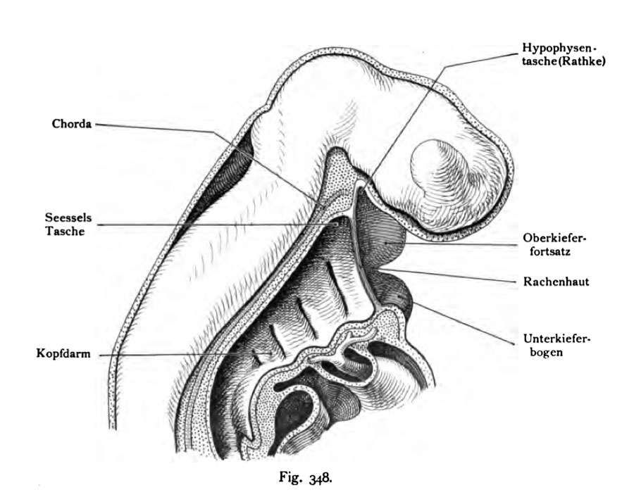
Fig. 348. The Pharyngeal Membrane of the human embryo marked by a dotted line in the sagittal section shown in the head
Embryo of 4.2 mm in length. Age 31 - 34 days.
At this time the pharyngeal membrane is already torn throat. But she was here- given to their construction from ectoderm and endoderm, and its location to Rathke's between S and eat Ischen present case. The upper boundary between mouth bay and foregut in later stages of development identified, e.g. in Figures 355 and 356 and with the adults in the front half So in the sella turcica region of parasphenoid. The extension on the ground the oral cavity is the 348 on FIG free dorsal edge of the mandibular continuous rate. In abnormal cases can persist for pharyngeal membrane in mouth bay and throat wear red skin colour.
Rachenhaut = Pharyngeal Membrane
- This text is a Google translate computer generated translation and may contain many errors.
Images from - Atlas of the Development of Man (Volume 2)
(Handatlas der entwicklungsgeschichte des menschen)
- Kollmann Atlas 2: Gastrointestinal | Respiratory | Urogenital | Cardiovascular | Neural | Integumentary | Smell | Vision | Hearing | Kollmann Atlas 1 | Kollmann Atlas 2 | Julius Kollmann
- Links: Julius Kollman | Atlas Vol.1 | Atlas Vol.2 | Embryology History
| Historic Disclaimer - information about historic embryology pages |
|---|
 |
Reference
Kollmann JKE. Atlas of the Development of Man (Handatlas der entwicklungsgeschichte des menschen). (1907) Vol.1 and Vol. 2. Jena, Gustav Fischer. (1898).
Cite this page: Hill, M.A. (2024, April 26) Embryology Kollmann348.jpg. Retrieved from https://embryology.med.unsw.edu.au/embryology/index.php/File:Kollmann348.jpg
- © Dr Mark Hill 2024, UNSW Embryology ISBN: 978 0 7334 2609 4 - UNSW CRICOS Provider Code No. 00098G
Fig. 348. Die Rachenhaut des menschlichen Embryo
durch eine punktierte Linie in den im Sagittalschnitt dargestellten Kopf eingezeichnet.
Embryo von 4,2 mm Länge. Alter 31 - 34 Tage.
Um diese Zeit ist die Rachenhaut bereits eingerissen. Sie wurde aber hier an- gegeben, um ihren Bau aus Ekto- und Entoderm und ihre Lage zur Rathke- schen und S esse Ischen Tasche darzustellen. Die obere Grenze zwischen Mundbucht und Kopfdarm in allen späteren Entwicklungsstufen erkennbar, z. B. in den Fig. 355 und 356 und bei dem Erwachsenen an der vorderen Hälfte der Sella turcica also im Gebiet des Präsphenoid. Die Ausdehnung am Boden der Mundhöhle liegt in der Fig. 348 am freien dorsalen Rande des Unterkiefer- fortsatzes. In abnormen Fällen kann die Rachenhaut persistieren. Mundbucht und Rachenhaut tragen rote Farbe.
File history
Click on a date/time to view the file as it appeared at that time.
| Date/Time | Thumbnail | Dimensions | User | Comment | |
|---|---|---|---|---|---|
| current | 12:56, 16 October 2011 | | 883 × 706 (87 KB) | S8600021 (talk | contribs) | {{Kollmann1907}} |
You cannot overwrite this file.
File usage
The following page uses this file: