File:Journal.pone.0013559.g001.png
Journal.pone.0013559.g001.png (529 × 600 pixels, file size: 501 KB, MIME type: image/png)
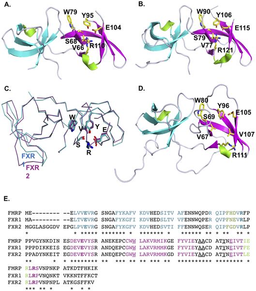
Tud 1 domains are colored in cyan and Tud2 domains in magenta. Coiled regions are indicated in grey. The residues forming the aromatic cage of Tud2 are shown as in stick representation and are colored yellow. (C) FXR1 (cyan) and FXR2 (purple) align well and reveal a conserved interdomain orientation. (D The previously determined structure of FMRP (PDB 2BDK) also comprises the tandem Tudor architecture. The coloring is as described for the FXR1 and FXR2 panels. (E) The sequence alignment of the FXR proteins. Residues are colored in agreement with the β-strands of panels A, B, and D. Residues in bold correspond to the ionic lock, underlined residues exhibit alterations in the HSQC spectra on peptide titration, and the asterisks denote strictly conserved residues.
http://www.plosone.org/article/info%3Adoi%2F10.1371%2Fjournal.pone.0013559
Copyright: © 2010 Adams-Cioaba et al. This is an open-access article distributed under the terms of the Creative Commons Attribution License, which permits unrestricted use, distribution, and reproduction in any medium, provided the original author and source are credited.
--Boris Zolotarev 10:52, 18 August 2011 (EST)
File history
Click on a date/time to view the file as it appeared at that time.
| Date/Time | Thumbnail | Dimensions | User | Comment | |
|---|---|---|---|---|---|
| current | 10:47, 18 August 2011 | | 529 × 600 (501 KB) | Z3290689 (talk | contribs) | Tud 1 domains are colored in cyan and Tud2 domains in magenta. Coiled regions are indicated in grey. The residues forming the aromatic cage of Tud2 are shown as in stick representation and are colored yellow. (C) FXR1 (cyan) and FXR2 (purple) align well a |
You cannot overwrite this file.
File usage
The following page uses this file: