File:Jimenez-Castellanos1949 fig04.jpg

Jimenez-Castellanos1949_fig04.jpg (728 × 521 pixels, file size: 78 KB, MIME type: image/jpeg)
Fig. 4. Embryo 9 mm
Photomicrograph through a human embryo of 9 mm. at the level indicated in fig. 1. :1: 59. (1) Aorta; (2) posterior cardinal vein; (3) mesonephros; (4) gonad; (5) liver; (6) juxtaaortic body; (7) body of vertebra.
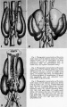
Fig 1-3
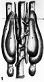
Fig 1
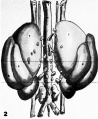
Fig 2
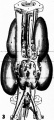
Fig 3
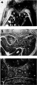
Fig 4-6
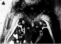
Fig 4
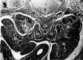
Fig 5
Fig 6


Reference
Jimenez-Castellanos J. The morphogenesis of the systems of juxta-aortic tissues in human embryos. (1949) Q Bull Northwest Univ Med Sch. 23(4):428-31. PMID: 18148736
Cite this page: Hill, M.A. (2024, April 26) Embryology Jimenez-Castellanos1949 fig04.jpg. Retrieved from https://embryology.med.unsw.edu.au/embryology/index.php/File:Jimenez-Castellanos1949_fig04.jpg
- © Dr Mark Hill 2024, UNSW Embryology ISBN: 978 0 7334 2609 4 - UNSW CRICOS Provider Code No. 00098G
File history
Click on a date/time to view the file as it appeared at that time.
| Date/Time | Thumbnail | Dimensions | User | Comment | |
|---|---|---|---|---|---|
| current | 04:03, 17 August 2017 | | 728 × 521 (78 KB) | Z8600021 (talk | contribs) |
You cannot overwrite this file.
File usage
The following 9 pages use this file:
- Paper - The morphogenesis of the systems of juxta-aortic tissues in human embryos
- File:Jimenez-Castellanos1949 fig01-3.jpg
- File:Jimenez-Castellanos1949 fig01.jpg
- File:Jimenez-Castellanos1949 fig02.jpg
- File:Jimenez-Castellanos1949 fig03.jpg
- File:Jimenez-Castellanos1949 fig04-6.jpg
- File:Jimenez-Castellanos1949 fig04.jpg
- File:Jimenez-Castellanos1949 fig05.jpg
- File:Jimenez-Castellanos1949 fig06.jpg