File:Ingalls1932b plate99.jpg

Original file (862 × 1,320 pixels, file size: 192 KB, MIME type: image/jpeg)
Plate 99
Fig. 1. Embryo No. 83. Greatest length about 7mm. Open neural tube in sacral region. Facial features distorted, heart exposed.
Fig. 2. Embryo No. 46. Greatest length 14.5mm. The entire body is very much malformed. Almost the whole of the dorsum is markedly altered or defective. The deep triangular cavity is due to the postmortem loss of tissue.
Fig. 3. Embryo No. 129. Greatest length 12mm., dorsal view. Irregular, roughly circular discolored area in the anterior part of the back. Entire body badly stunted and deformed.
Fig. 4. Embryo No. 161. Greatest length about 12mm. Symmetrically located, transversely elongated area over the anterior part of the rhombencephalon. Embryo in very poor condition.
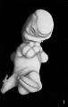
Fig. 1. Embryo No. 83.
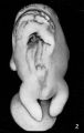
Fig. 2. Embryo No. 46.
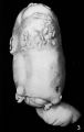
Fig. 3. Embryo No. 129.
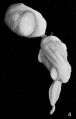
Fig. 4. Embryo No. 161.


Reference
Ingalls NW. Studies in the pathology of development: II. Some aspects of defective development in the dorsal midline. (1932) Am J Pathol. 8(5): 525-556 PMID 19970035
Cite this page: Hill, M.A. (2024, April 26) Embryology Ingalls1932b plate99.jpg. Retrieved from https://embryology.med.unsw.edu.au/embryology/index.php/File:Ingalls1932b_plate99.jpg
- © Dr Mark Hill 2024, UNSW Embryology ISBN: 978 0 7334 2609 4 - UNSW CRICOS Provider Code No. 00098G
File history
Click on a date/time to view the file as it appeared at that time.
| Date/Time | Thumbnail | Dimensions | User | Comment | |
|---|---|---|---|---|---|
| current | 08:53, 14 October 2020 | | 862 × 1,320 (192 KB) | Z8600021 (talk | contribs) | ===Reference=== {{Ref-Ingalls1932b}} {{footer}} |
You cannot overwrite this file.
File usage
The following page uses this file: