File:Ingalls1932b plate101.jpg
Original file (857 × 1,308 pixels, file size: 174 KB, MIME type: image/jpeg)
Plate 101
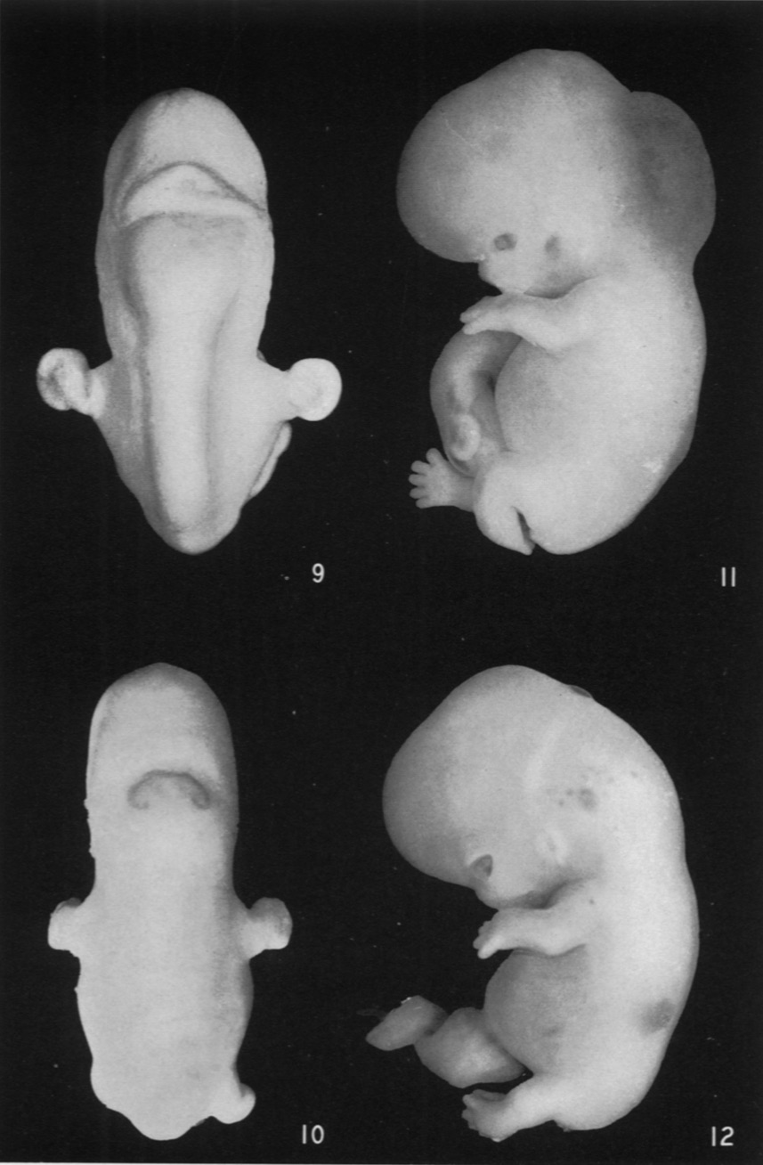
Fig. 9. Embryo No. 652. Greatest length 15 mm. Dorsal view of anterior end of embryo to show the transverse discolored band just behind the midbrain.
Fig. 10. Embryo No. 167. Greatest length 18.5mm. Very conspicuous, sharply defined, discolored area over the rhombencephalon. There is a similar smaller patch over the vertex and two paired spots on the upper part of the forehead.
Fig. 11. Embryo No. 671. Greatest length 25 mm. Enormous blood-stained bleb on back of head and neck. Jn the lower thoracic region there is a much smaller one. Small ecchymoses on face and arm.
Fig. 12. Embryo No. 611. Greatest length 23 mm. Minute, translucent bleb over the cerebellum. Ecchymoses on head, shoulder and trunk.
Fig. 9. Embryo No. 652.
Fig. 10. Embryo No. 167.
Fig. 11. Embryo No. 671.
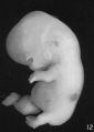
Fig. 12. Embryo No. 611.


Reference
Ingalls NW. Studies in the pathology of development: II. Some aspects of defective development in the dorsal midline. (1932) Am J Pathol. 8(5): 525-556 PMID 19970035
Cite this page: Hill, M.A. (2024, April 27) Embryology Ingalls1932b plate101.jpg. Retrieved from https://embryology.med.unsw.edu.au/embryology/index.php/File:Ingalls1932b_plate101.jpg
- © Dr Mark Hill 2024, UNSW Embryology ISBN: 978 0 7334 2609 4 - UNSW CRICOS Provider Code No. 00098G
File history
Click on a date/time to view the file as it appeared at that time.
| Date/Time | Thumbnail | Dimensions | User | Comment | |
|---|---|---|---|---|---|
| current | 08:53, 14 October 2020 | | 857 × 1,308 (174 KB) | Z8600021 (talk | contribs) | ===Reference=== {{Ref-Ingalls1932b}} {{footer}} |
You cannot overwrite this file.
File usage
The following page uses this file: