File:Ingalls1932b plate100.jpg
Original file (863 × 1,329 pixels, file size: 197 KB, MIME type: image/jpeg)
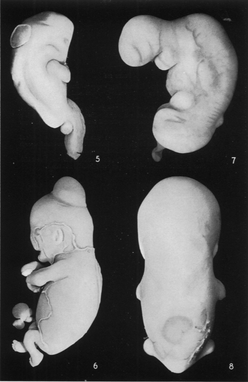
Plate 100
Fig. 5. Embryo No. 210. Greatest length 15mm. Large thin-walled bleb in the midline over the rhombencephalon. Much deformity in the body generally.
Fig. 6. Embryo No. 597 B. The smaller of a pair of binoval twins, greatest length 32.5mm. Very large, rather thick-walled bleb in the midline of head, just behind vertex. Extensive desquamation, malformed hands and cord.
Fig. 7. Embryo No. 407. Greatest length 7 mm. Small bleb-like elevation of epithelium in the midline of the back. Head small and malformed.
Fig. 8. Embryo No. 442. Greatest length 26 mm. Dark patch in lower dorsal region; on the right and below can be seen the borders of the larger area. Laterally the epidermal thickenings are very conspicuous. There is also a very definite transverse band across the forehead.
Fig. 5. Embryo No. 210.
Fig. 6. Embryo No. 597 B.
Fig. 7. Embryo No. 407.
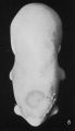
Fig. 8. Embryo No. 442.


Reference
Ingalls NW. Studies in the pathology of development: II. Some aspects of defective development in the dorsal midline. (1932) Am J Pathol. 8(5): 525-556 PMID 19970035
Cite this page: Hill, M.A. (2024, April 26) Embryology Ingalls1932b plate100.jpg. Retrieved from https://embryology.med.unsw.edu.au/embryology/index.php/File:Ingalls1932b_plate100.jpg
- © Dr Mark Hill 2024, UNSW Embryology ISBN: 978 0 7334 2609 4 - UNSW CRICOS Provider Code No. 00098G
File history
Click on a date/time to view the file as it appeared at that time.
| Date/Time | Thumbnail | Dimensions | User | Comment | |
|---|---|---|---|---|---|
| current | 08:53, 14 October 2020 | | 863 × 1,329 (197 KB) | Z8600021 (talk | contribs) | ===Reference=== {{Ref-Ingalls1932b}} {{footer}} |
You cannot overwrite this file.
File usage
The following page uses this file: