File:Ingalls1918 plate 2 fig5+6.jpg
Original file (1,000 × 356 pixels, file size: 60 KB, MIME type: image/jpeg)
Plate 2
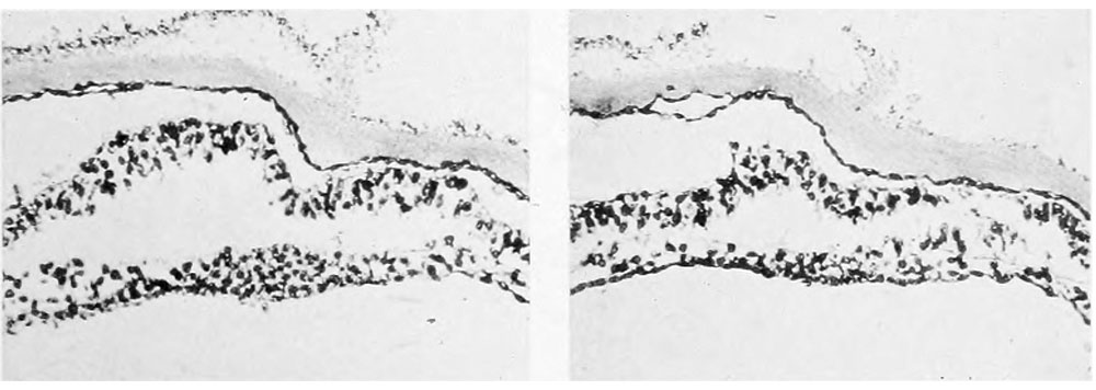
Photographs of sections of axial structures, the location and direction of which are indicated in the text-figures. In all the photographs and drawings the right side of the embryo is on the left in the plates, i. e. all views looking caudad. Dorsal structures are cut slightly more anterior than ventral ones. (Cf. text-figure 3.)
1. Section 425. Posterior part of primitive streak. X 160.
2. Section 406. Most caudal section in which archenteric canal appears. Its location may be recognized by the absence of nuclei. X 160.
3. Section 401. Archenteric canal where largest and be.st defined. X 400.
4. Section 395. Large (second) ventral opening, "plaque notochordale" very distinct. X 400.
5. Section 380. Completion plate where best developed. X 160.
6. Section S75. Completion plate near anterior limit. X 160.
- Contribution No.23: Figures | Plate 1 | Plate 2 | Plate 3 | Plate 4 | Plate 1 | Carnegie - Contributions to Embryology | Carnegie stage 8 | Category:Carnegie Stage 8 | Historic Embryology Papers
| Historic Disclaimer - information about historic embryology pages |
|---|
 |
Reference
Ingalls NW. A human embryo before the appearance of the myotomes. (1918) Contrib. Embryol., Carnegie Inst. Wash. No.23 Publ. 227, 7:111-134.
Cite this page: Hill, M.A. (2024, April 26) Embryology Ingalls1918 plate 2 fig5+6.jpg. Retrieved from https://embryology.med.unsw.edu.au/embryology/index.php/File:Ingalls1918_plate_2_fig5%2B6.jpg
- © Dr Mark Hill 2024, UNSW Embryology ISBN: 978 0 7334 2609 4 - UNSW CRICOS Provider Code No. 00098G
| Historic Disclaimer - information about historic embryology pages |
|---|
|
File history
Click on a date/time to view the file as it appeared at that time.
| Date/Time | Thumbnail | Dimensions | User | Comment | |
|---|---|---|---|---|---|
| current | 11:40, 2 January 2013 | 1,000 × 356 (60 KB) | Z8600021 (talk | contribs) | ===Plate 2=== Photographs of sections of axial structures, the location and direction of which are indicated in the text-figures. In all the photographs and drawings the right side of the embryo is on the left in the plates, i. e. all views looking cauda |
You cannot overwrite this file.
File usage
The following page uses this file: