File:Human fetal adrenal gland 01.jpg

Original file (1,266 × 800 pixels, file size: 107 KB, MIME type: image/jpeg)
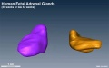
Human Fetal Adrenal Gland 3D Models
| The fetal adrenal gland appears as a pyramidal or half-moon shape, and almost remains the same during 23–40 weeks GA.
The medial surface is flat. The groove on the lateral surface becomes shallower as GA increases. The bar in each figure represents 1 centimeter.
|
|
Reference
Zhang Z, Meng H, Hou Z, Ma J, Feng L, Lin X, Tang Y, Zhang X, Liu Q & Liu S. (2013). Fetal adrenal gland in the second half of gestation: morphometrical assessment with 3.0T post-mortem MRI. PLoS ONE , 8, e75511. PMID: 24116052 DOI.
Copyright
This is an open-access article distributed under the terms of the Creative Commons Attribution License, which permits unrestricted use, distribution, and reproduction in any medium, provided the original author and source are credited.
Figure 4. Journal.pone.0075511.g004.jpg Original figure adjusted in size, sharpness and labelling.
Cite this page: Hill, M.A. (2024, April 26) Embryology Human fetal adrenal gland 01.jpg. Retrieved from https://embryology.med.unsw.edu.au/embryology/index.php/File:Human_fetal_adrenal_gland_01.jpg
- © Dr Mark Hill 2024, UNSW Embryology ISBN: 978 0 7334 2609 4 - UNSW CRICOS Provider Code No. 00098G
File history
Click on a date/time to view the file as it appeared at that time.
| Date/Time | Thumbnail | Dimensions | User | Comment | |
|---|---|---|---|---|---|
| current | 15:55, 25 November 2014 | | 1,266 × 800 (107 KB) | Z8600021 (talk | contribs) | ==Human Fetal adrenal gland 3D models== They are 23 (A), 27(B), 32(C), and 36 (D) weeks GA, respectively. The fetal adrenal gland appears as a pyramidal or half-moon shape, and almost remains the same during 23–40 weeks GA. The medial surface is fl... |
You cannot overwrite this file.
File usage
The following page uses this file: