File:Human embryo midgut loop 01.jpg
Original file (1,423 × 771 pixels, file size: 200 KB, MIME type: image/jpeg)
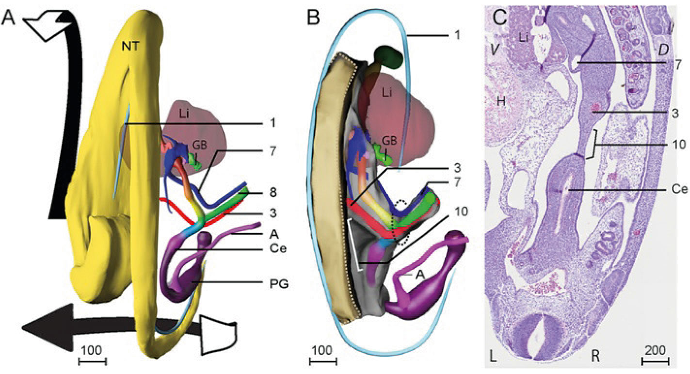
Human Embryo Midgut Loop
The orientation of the midgut loop and its mesentery during the 5th week follows the helical body axis.
- A - Dorsal view of the reconstruction of a CS14 embryo (s5029). Note the left-sided juxtaposition of the head relative to the caudal end of the body, reflecting the helical body axis. The successive parts of the midgut are shown in a rainbow color gradient (see legend color codes). Note that the vitelline artery (3) and the right vitelline vein (7) traverse the vitelline duct (8) at the apex of the midgut loop. The arrows indicate the changes occurring during straightening of the body axis in CS15 and CS16 embryos.
- B - shows the position of the developing midgut mesentery (10) between both limbs of the midgut. Note the limited craniocaudal extension of the mesentery at this stage (10). The beige area identifies the region where the intestinal mesenchyme is attached to the dorsal body wall.
- C - Histological section of embryo s5029 with right vitelline vein (7), vitelline artery (3), cecum (Ce) and developing dorsal midgut mesentery (10). Due to the helical body axis, the caudal end of the body is cut near transversely (with left (L) and right (R) sides), whereas more cranially, the body is cut almost sagittally (V: ventral; D: dorsal). Note that the midgut mesentery (10) is ~4-fold thinner than the mesenchymal mass surrounding the intestine.
Scale bar unit: μm.
- Links: week 5 | secondary loops week 7 to 8 | tertiary loops week 8 | Intestine Development | Week 7 | Week 8
Reference
<pubmed>26297675</pubmed>
https://bmcdevbiol.biomedcentral.com/articles/10.1186/s12861-015-0081-x
Copyright
© 2015 Soffers et al. Open Access This article is distributed under the terms of the Creative Commons Attribution 4.0 International License (http://creativecommons.org/licenses/by/4.0), which permits unrestricted use, distribution, and reproduction in any medium, provided you give appropriate credit to the original author(s) and the source, provide a link to the Creative Commons license, and indicate if changes were made. The Creative Commons Public Domain Dedication waiver (http://creativecommons.org/publicdomain/zero/1.0/) applies to the data made available in this article, unless otherwise stated.
http://www.biomedcentral.com/1471-213X/15/31/figure/F1
S12861-015-0081-x-2.jpg
File history
Click on a date/time to view the file as it appeared at that time.
| Date/Time | Thumbnail | Dimensions | User | Comment | |
|---|---|---|---|---|---|
| current | 20:54, 24 August 2015 | | 1,423 × 771 (200 KB) | Z8600021 (talk | contribs) | ==Human Embryo Midgut Loop)== The orientation of the midgut loop and its mesentery during the 5th week follows the helical body axis. * '''A''' - Dorsal view of the reconstruction of a CS14 embryo (s5029). Note the left-sided juxtaposition of the he... |
You cannot overwrite this file.
File usage
The following page uses this file: