File:Human Embryo 17.8mm GIT.jpg
Human_Embryo_17.8mm_GIT.jpg (600 × 401 pixels, file size: 38 KB, MIME type: image/jpeg)
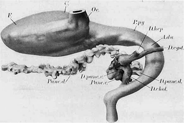
Human Embryo (17.8mm) Gastrointestinal Tract
Wax-plate reconstruction of the stomach, the duodenum and the pancreas. The model is represented somewhat ventrally, from the right side.
- A. du., antrum duodenale
- C., corpus gastri
- D. chol., ductus choledochus
- D. cysl.. ductus cysticus
- D. hep., ductus hepaticus
- D. panc d., ductus pancreatis dorsalis,
- D. panc. E., ductus pancreatis ventralis
- F., fundus gastri
- Oe., oesophagus
- Panr d , pancreas dorsale
- Pam. v., pancreas ventrale
- P. py , pars pylorica pastri
Online Editor Notes
- The embryo external appearance and dimensions suggest that it is a Carnegie stage 19 embryo (Week 7, 48 - 51 days, 16 - 18 mm).
- This figure appeared in 1908 as Fig. 6. Reconstruction from a human embryo of 13.6 mm
Reference
Thyng FW. The anatomy of a 17.8 mm human embryo. (1914) Amer. J Anat. 17: 31-112.
A reproduction of figure 104, page 153 of “Laboratory Textbook of Embryology,” Charles Sedgwick Minot, edition of 1910, published by P. Blakiston’s Son and Company, Philadelphia. The American Journal of Anatomy, Vol.17, No.1 These drawings are based on studies of the Harvard Embryological Collection while he was in Minot's Lab in 1907-08. He was also an anatomist at Northwestern University Medical School.
Cite this page: Hill, M.A. (2024, April 26) Embryology Human Embryo 17.8mm GIT.jpg. Retrieved from https://embryology.med.unsw.edu.au/embryology/index.php/File:Human_Embryo_17.8mm_GIT.jpg
- © Dr Mark Hill 2024, UNSW Embryology ISBN: 978 0 7334 2609 4 - UNSW CRICOS Provider Code No. 00098G
File history
Click on a date/time to view the file as it appeared at that time.
| Date/Time | Thumbnail | Dimensions | User | Comment | |
|---|---|---|---|---|---|
| current | 13:29, 4 August 2009 |  | 600 × 401 (38 KB) | MarkHill (talk | contribs) | Human Embryo (17.8mm) Gastrointestinal Tract Wax-plate reconstruction of the stomach, the duodenum and the pancreas. The model is represented somewhat ventrally, from the right side A. du., antrum duodenale; C., corpus gastri; D. chol., ductus choledoch |
You cannot overwrite this file.
File usage
The following 4 pages use this file:
