File:Holyoke1936 plate 02.jpg

Original file (1,000 × 1,504 pixels, file size: 250 KB, MIME type: image/jpeg)
Plate 2
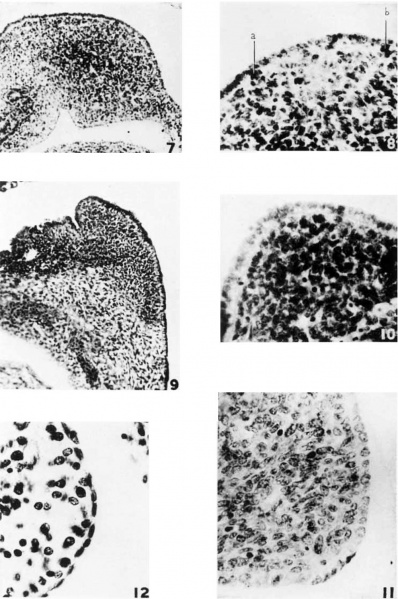
7 Pig embryo, 14 mm., showing the forination of the splenie fold in the dorsal niesogastrium and the bulging due to the growth of the anlage. The splenic condensation is distinct from the surrounding niesencliyiiio, particularly in the region of the splenic vessels.
8 Detail froin preceding figure, showing the euhoida.l eoelomie epitheliuin and the early formation of the light zone. 4 and 1) indicate cells passing into the mesenchyme. Photo X 550.
9 Huinan embryo, 15 mm. The spleen is situated in the convexity of a sharp bend in the transvse.11tory. The hilus is located in the loose iricsenehyme in the eoiieavity of this bend. The eoelomie epithelium is a columnar layer completely out off from the spleen by a basement nnenibrane. Hematoxylin and orange G. Plmto X 200.
10 Pig embryo, 22 mm. There is 4, distinct euboidal layer of aoeloniic. epithelium sharply mzu'ked off by a basement membrane. Delafield’s heamtoxylin and azure II eosin. Photo X 550.
11 Pig embryo, 35 mm., showing the persistence of a euboidal type of mesotheliuin over the spleen. Delafield’s hematoxylin and azure II eosin. photo X 750.
12 Human embryo, 22 mm. There is a distinct low euhoidal layer of epithelium simulating somewhat the adult, condition. Ilexrmtoxyliii mid eosin. Photo X 750.
| Historic Disclaimer - information about historic embryology pages |
|---|
| Pages where the terms "Historic" (textbooks, papers, people, recommendations) appear on this site, and sections within pages where this disclaimer appears, indicate that the content and scientific understanding are specific to the time of publication. This means that while some scientific descriptions are still accurate, the terminology and interpretation of the developmental mechanisms reflect the understanding at the time of original publication and those of the preceding periods, these terms, interpretations and recommendations may not reflect our current scientific understanding. (More? Embryology History | Historic Embryology Papers) |
Reference
Holyoke EA. The role of the primitive mesothelium in the development of the mammalian spleen. (1936) Anat. Rec. 65(3): 333-349.
Cite this page: Hill, M.A. (2024, April 27) Embryology Holyoke1936 plate 02.jpg. Retrieved from https://embryology.med.unsw.edu.au/embryology/index.php/File:Holyoke1936_plate_02.jpg
- © Dr Mark Hill 2024, UNSW Embryology ISBN: 978 0 7334 2609 4 - UNSW CRICOS Provider Code No. 00098G
File history
Click on a date/time to view the file as it appeared at that time.
| Date/Time | Thumbnail | Dimensions | User | Comment | |
|---|---|---|---|---|---|
| current | 16:55, 20 March 2017 |  | 1,000 × 1,504 (250 KB) | Z8600021 (talk | contribs) | |
| 16:55, 20 March 2017 |  | 1,603 × 2,428 (443 KB) | Z8600021 (talk | contribs) | {{Historic Disclaimer}} ===Reference=== {{Ref-Holyoke1936}} {{Footer}} Category:Spleen |
You cannot overwrite this file.
File usage
The following page uses this file:
