File:His1897 plate01.jpg

Original file (1,798 × 2,877 pixels, file size: 745 KB, MIME type: image/jpeg)
Plate I
Fig 7. Mesial longitudinal section through the neural tube of an early chick embryo. The aperture or neuropore (n.p.) at the anterior end of the tube is still open.
Fig. 8. Mesial longitudinal section through the head-end of a slightly older chick embryo, showing the intimate connection between the anterior end of the notochord (n.) and the hinder part of the floor of the fore-brain. Note that the neuropore is closed.
Fig. 9. Early embryo of a dog-fish. The upper end of the figure shows ie enlargement and bending of the brain-part of the neural tube.
Fig. 10. Micropliotograph of a portion. of a transverse section through the neural tube. The inner ends of the spongioblastic columns are observed to be flattened out and joined together to form a fine limiting membrane. This is seen in the upper part of the section. Between them three germinal cells, two on the left with karyokinetic formations, may be seen. - Human embryo of four weeks.
Fig. 11. In this microphotograph the neuroblastic cells are darkcoloured, and in connection with many of these the tapering axis cylinder processes are seen. It will be observed that these processes are growing towards the surface ( i.e the lower part of the figure) through the mesh-work of the myelosponge. - Human embryo of four weeks.
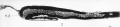
Fig 7. Mesial longitudinal section through the neural tube of an early chick embryo. The aperture or neuropore (n.p.) at the anterior end of the tube is still open.
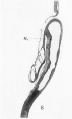
Fig. 8. Mesial longitudinal section through the head-end of a slightly older chick embryo, showing the intimate connection between the anterior end of the notochord (n.) and the hinder part of the floor of the fore-brain. Note that the neuropore is closed.
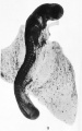
Fig. 9. Early embryo of a dog-fish. The upper end of the figure shows ie enlargement and bending of the brain-part of the neural tube.
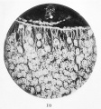
Fig. 10. Micropliotograph of a portion of a transverse section through the neural tube. The inner ends of the spongioblastic columns are observed to be flattened out and joined together to form a fine limiting membrane. This is seen in the upper part of the section. Between them three germinal cells, two on the left with karyokinetic formations, may be seen. Human embryo of four weeks.
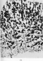
Fig. 11. In this microphotograph the neuroblastic cells are darkcoloured, and in connection with many of these the tapering axis cylinder processes are seen. It will be observed that these processes are growing towards the surface ( i.e the lower part of the figure) through the mesh-work of the myelosponge. Human embryo of four weeks.


| Historic Disclaimer - information about historic embryology pages |
|---|
 |
- Links: Fig 1 | Fig 2 | Fig 3 | Fig 4 | Fig 5 | Fig 6 | Plate 1 | Fig 7 | Fig 8 | Fig 9 | Fig 10 | Fig 11 | Plate 2 | Fig 12 | Fig 13 | Fig 14 | Fig 15 | Plate 3 | Fig 16 | Fig 17 | Plate 4 | Fig 18 | Fig 19 | Fig 20 | Fig 21 | Fig 22 | Fig 23 | Plate 5 | Fig 24 | Fig 25 | Fig 26 | Fig 27 | Fig 28 | His 1897 | Wilhelm His | Historic Embryology Papers
Reference
His W. Address upon the development of the brain. (1897) Trans. Royal Acad. Medicine Ireland.
Cite this page: Hill, M.A. (2024, April 26) Embryology His1897 plate01.jpg. Retrieved from https://embryology.med.unsw.edu.au/embryology/index.php/File:His1897_plate01.jpg
- © Dr Mark Hill 2024, UNSW Embryology ISBN: 978 0 7334 2609 4 - UNSW CRICOS Provider Code No. 00098G
File history
Click on a date/time to view the file as it appeared at that time.
| Date/Time | Thumbnail | Dimensions | User | Comment | |
|---|---|---|---|---|---|
| current | 19:10, 17 January 2016 | | 1,798 × 2,877 (745 KB) | Z8600021 (talk | contribs) |
You cannot overwrite this file.
File usage
The following page uses this file: