File:HillFlorian1931 text-fig02.jpg
Original file (1,672 × 2,111 pixels, file size: 276 KB, MIME type: image/jpeg)
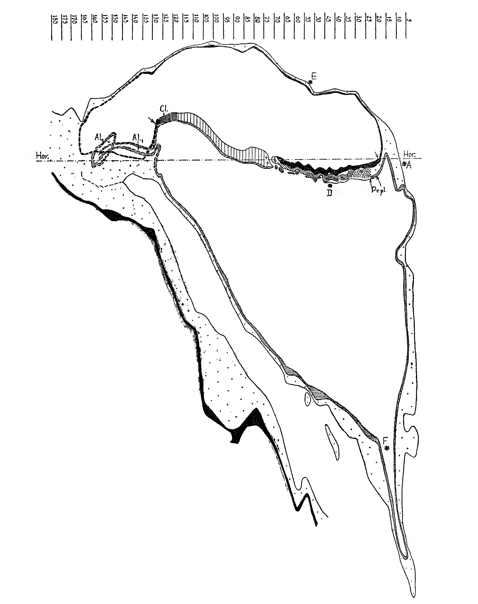
Text-Fig. 2. Graphic reconstruction of the median section through the Dobbin embryo
Ectoderm black; endoderm lined horizontally; primitive streak lined vertically; += HENSEN’s knot; chorda-process lined obliquely; prochordal plate (Pr.pl.) lined horizontally and dotted; mesoderm dotted; cloacal membrane (O'l.) cross—hatched. Limits of the embryonal shield marked by arrows. AL; 2: proximal (tubular) part of the allantoic canal; Al.2 = its distal (vesicular) part.
Hor. —-—=. horizontal plane (the plane of the projection in text-figs. 1 and 8). The caudal part of the embryonic formation, including the cloacal membrane, the allantoic canal, the caudal part of the amniotic cavity and the connecting stalk are projected into the median plane, and their outlines are marked by interrupted lines. , The cell-layer underneath the intermediate region and the most cranial portion of the head-process, the significance of Which is doubtful, is left White. X 75.
| Anterior margin, embryonal shield to region of cloacal membrane (A-Cl.) | 0.98 mm |
| Vertical diameter (D-F) of yolk-sac | 1.092 mm |
| Antero-posterior diameter of yolk-sac, near its mid-region | 0.98 mm |
| Vertical height, amnio-embryonal vesicle (D-E) | 0.468 mm |
| Length of yolk-sac process | 0.88 mm |
| Historic Disclaimer - information about historic embryology pages |
|---|
 |
Reference
Hill JP. and Florian J. A young human embryo (embryo dobbin) with head-process and prochordal plate. (1931) Phil. Tran. Roy. Soc. London B, 219: 443-486.
Cite this page: Hill, M.A. (2024, April 27) Embryology HillFlorian1931 text-fig02.jpg. Retrieved from https://embryology.med.unsw.edu.au/embryology/index.php/File:HillFlorian1931_text-fig02.jpg
- © Dr Mark Hill 2024, UNSW Embryology ISBN: 978 0 7334 2609 4 - UNSW CRICOS Provider Code No. 00098G
File history
Click on a date/time to view the file as it appeared at that time.
| Date/Time | Thumbnail | Dimensions | User | Comment | |
|---|---|---|---|---|---|
| current | 16:42, 10 August 2015 | | 1,672 × 2,111 (276 KB) | Z8600021 (talk | contribs) | |
| 16:41, 10 August 2015 | 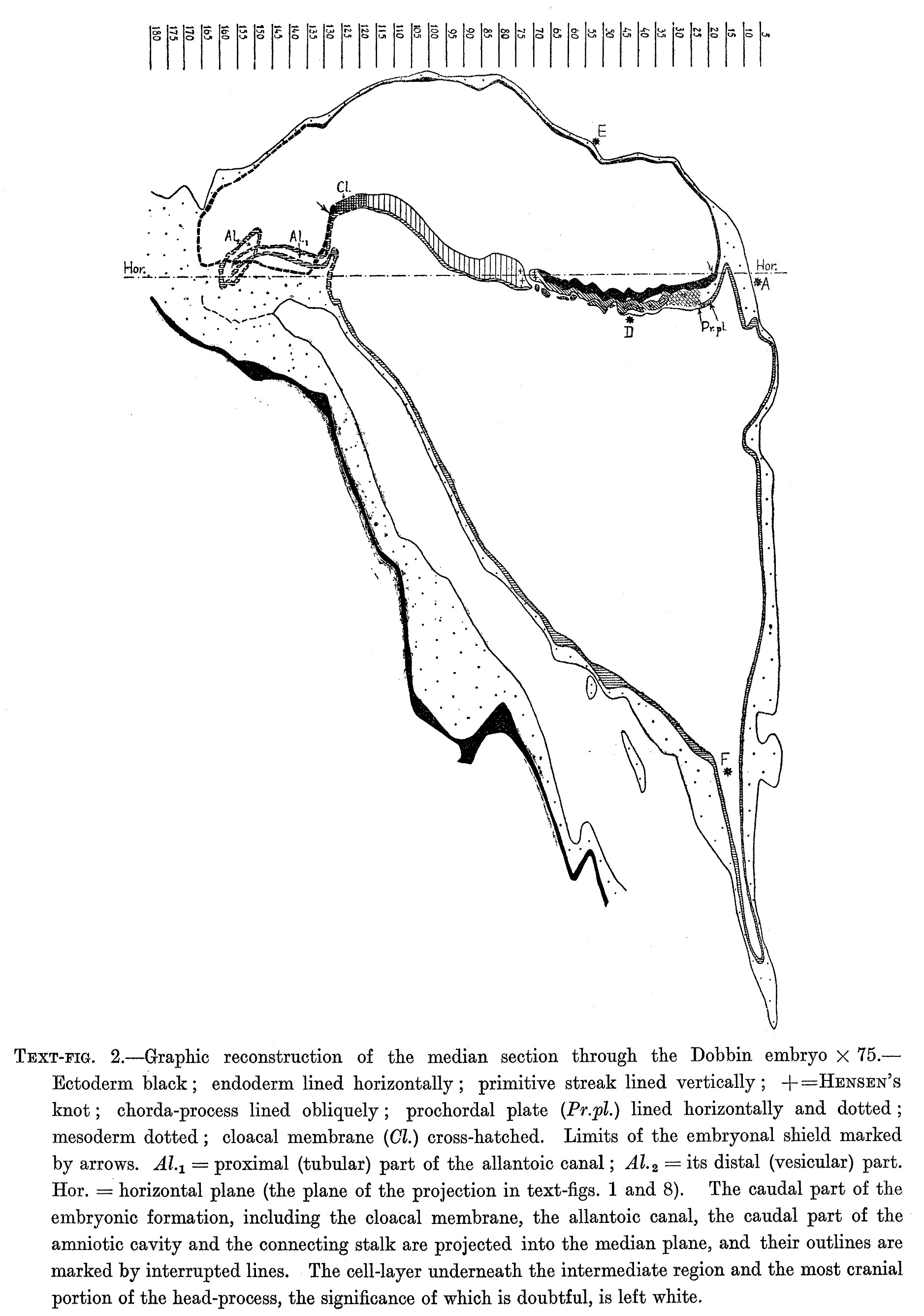 | 1,877 × 2,671 (547 KB) | Z8600021 (talk | contribs) |
You cannot overwrite this file.
File usage
The following 2 pages use this file: