File:Hertwig1892 fig320.jpg
From Embryology

Size of this preview: 706 × 600 pixels. Other resolution: 970 × 824 pixels.
Original file (970 × 824 pixels, file size: 179 KB, MIME type: image/jpeg)
Fig. 320. Sagittal reconstruction of a human embryo 5 mm long
neck measurement (embryo R, His), to illustrate the development of the pericardio-thoracic cavity and the diaphragm, after Wilhelm His (1831-1904).
ab, Aortic bulb ; brh, thoracic cavity (recessus parietalis, His) ; kh, pericardial cavity ; tie, ductus Cuvieri ; dc, vitelline vein (v. omphalomesenterica) ; nv, umbilical vein ; vca, cardinal vein ; vj, jugular vein ; Ig, lung ; z + I, fundament of the diaphragm and the liver ; uk, lower jaw.
| Historic Disclaimer - information about historic embryology pages |
|---|
 |
Reference
Historic Textbook Text-Book of the Embryology of Man and Mammals by Dr Oscar Hertwig (1892)
File history
Click on a date/time to view the file as it appeared at that time.
| Date/Time | Thumbnail | Dimensions | User | Comment | |
|---|---|---|---|---|---|
| current | 15:38, 21 February 2015 | | 970 × 824 (179 KB) | Z8600021 (talk | contribs) | |
| 15:38, 21 February 2015 | 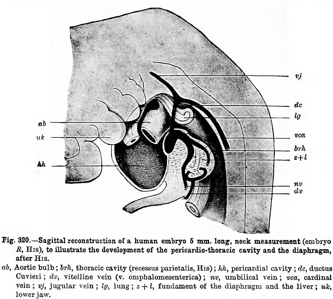 | 1,156 × 1,038 (260 KB) | Z8600021 (talk | contribs) | {{Hertwig1892 figures17-1}} |
You cannot overwrite this file.
File usage
The following page uses this file: