File:Hertig1956 fig88.jpg

Original file (722 × 780 pixels, file size: 51 KB, MIME type: image/jpeg)
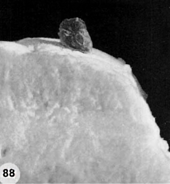
Plate 17. 12-day Previllous Ovum
The implantation site of a 12-day previllous ovum, normal except for a shallow type of implantation.
88 Profile view of the intact implantation site to show that the excessive hemorrhage completely conceals the shallowly implanted ovum seen in figures 89 and 90. Carnegie 8000, Sequence 3. X 5.
- Figure Links: 1 | 2 | 3 | 4 | 5 | 6 | 7 | 8 | 9-10 | 11-12 | 13-14 | 15-16 | 17 | 18-19 | 20 | 21-22 | 23 | 24-25 | 26-27 | 28-29 | 30-31 | 32-33 | 34 | 35 | 36 | 37 | 38 | 39 | 40 | 41 | 42 | 43 | 44 | 45 | 46 | 47 | 48 | 40 | 49 | 50 | 51 | 52 | 53 | 54 | 55 | 56 | 57 | 58 | 59 | 60 | 61 | 62 | 63 | 64 | 65 | 66 | 67 | 68 | 69 | 70 | 71 | 72 | 73 | 74 | 75 | 76 | 77 | 78 | 79 | 80 | 81 | 82 | 83 | 84 | 85 | 86 | 87 | 88 | 89 | 90 | plate 1 | plate 2 | plate 3 | plate 4 | plate 5 | plate 6 | plate 7 | plate 8 | plate 9 | plate 10 | plate 11 | plate 12 | plate 13 | plate 14 | plate 15 | plate 16 | plate 17 | table 1 | table 1 image | table 2 image | table 3 image | table 4 | table 4 image | table 5 | table 5 image | All figures | 1956 Hertig | Embryology History - Arthur Hertig | John Rock | Historic Papers
Reference
Hertig AT. Rock J. and Adams EC. A description of 34 human ova within the first 17 days of development. (1956) Amer. J Anat., 98:435-493.
Cite this page: Hill, M.A. (2024, May 1) Embryology Hertig1956 fig88.jpg. Retrieved from https://embryology.med.unsw.edu.au/embryology/index.php/File:Hertig1956_fig88.jpg
- © Dr Mark Hill 2024, UNSW Embryology ISBN: 978 0 7334 2609 4 - UNSW CRICOS Provider Code No. 00098G
File history
Click on a date/time to view the file as it appeared at that time.
| Date/Time | Thumbnail | Dimensions | User | Comment | |
|---|---|---|---|---|---|
| current | 23:24, 22 February 2017 | | 722 × 780 (51 KB) | Z8600021 (talk | contribs) | {{Hertig1956 figures}} |
You cannot overwrite this file.
File usage
The following 2 pages use this file: