File:Hertig1946b fig09.jpg
Original file (800 × 1,481 pixels, file size: 169 KB, MIME type: image/jpeg)
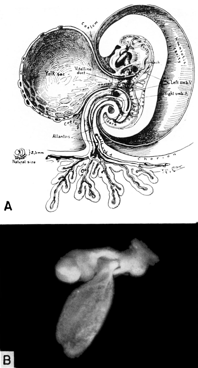
Fig. 9. A 2.5 mm and 3.2 mm embryo
A. A drawing of the right half of a 2.5 mm embryo representing a stage in the middle of the 4th week of development. The circulation is complete and the heart functions at this stage. Note the constriction of the yolk-sac to form the omphalomesenteric or vitelline duct as the digestive tract becomes more mature. The growth of the embryo and its surrounding amnion have resulted in the partial approximation of the body stalk and the vitelline duct so that these structures are being combined to form an early umbilical cord. (Fig. 3 from Cullen’s “The Umbilicus and Its Diseases,” W. B. Saunders Company.)
B. A 3.2 mm embryo with amnion removed as viewed from below and to the left showing the omphalo-mesenteric duct and its connection with yolk-sac and the primitive embryonic gut. Note the communication between the exocoelomic space around the yolk-sac and the coelom (body cavity) within the embryo. Carnegie 6488, sequence 2, X15. 104
References
Hertig AT. lnvolution of tissues in fetal life: a review. (1946) Anat. Rec. 94: 96-116.
{{Footer}
File history
Click on a date/time to view the file as it appeared at that time.
| Date/Time | Thumbnail | Dimensions | User | Comment | |
|---|---|---|---|---|---|
| current | 17:32, 7 August 2017 | | 800 × 1,481 (169 KB) | Z8600021 (talk | contribs) | ===References=== {{Ref-Hertig1946b}} {{Footer}} |
You cannot overwrite this file.
File usage
There are no pages that use this file.