File:Hertig1945d plate03.jpg

Original file (1,280 × 1,998 pixels, file size: 424 KB, MIME type: image/jpeg)
Plate 3. Ova of the twelfth and thirteenth days
Fig. 10. A mid-cross section of an 11.5-day ovum. The amniotic cavity is more advanced in its development than in the previous specimens although the amniogenic cells— now a distinct membrane - are still attached to the adjacent trophoblast. The exocoelome is now well contained within its membrane which is continuous above with the primitive endoderm of the germ-disk. Mesoblast formation is still continuing and is laying the foundation for the future connective tissue of the chorion. Carnegie 7699 section 8-5-3, X 100.
Fig. 11. The germ-disk, amnion and exocoeloomic membrane of the 11.5-day ovum at higher magnification to show greater detail. Note the attachment of the amniogenic cells to their parent trophoblast. Carnegie 7699, section 8-S-3, x 250.
Fig. 12. A mid-cross section of a 12.5-day ovum. The amniotic cavity is a cleft-like space above the primitive ectoderm of the germ-disk. It is barely enclosed above by the amniogenic cells which are still arising, in places, from adjacent trophoblast: Details of this process, at higher magnification, are shown in figs. 14 and 15. The exocoelomic (Heuser’s) membrane has reached the high point of its development and is destined to disintegrate within the next few days. Its essential mesoblastic nature is shown by the character of its cells and by their attachment. in places, to the trophoblast from which they arose. Carnegie 7700, section 6-1-S, x 100.
Fig. 13. plastic reconstruction of half the 12.5-day ovum. The cut surface represents the section seen in fig. 12. This picture, by its three dimensional concept serves to emphasize the nature of the amnion, the einocoelomic membrane and the mesoblast lining the chorionic cavity at this stage in their development. Carnegie 7700, reconstruction, x 75.
Fig. 14. A mid-cross section of the amnion and germ-disk of the 12.5-day ovum (same section as fig. 12). The cleft-like amniotic cavity is seen dorsal to the primitive ectoderm and is barely enclosed by the extremely thin amnion ab-ovve. Carnegie 7700, section 6-1-5, X 250.
Fig. 15. para-median section of the germ-disk and amniotic cavity from the 12.5-day ovum. Note the variable thickness and appearance of the amniogenic cells which are being added to by dividing cells derived from the adjacent trophoblast. Carnegie 7700, section S-7-7, x 250
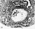
Fig 10 Carnegie 7699
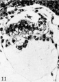
Fig 11 Carnegie 7699
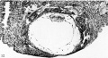
Fig 12 Carnegie 7700
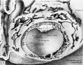
Fig 13 Carnegie 7700
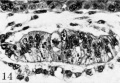
Fig 14 Carnegie 7700
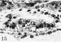
Fig 15 Carnegie 7700


Reference
Hertig AT. On the development of the amnion and exocoelomic membrane in the previllous human ovum. (1945) Yale J Biol Med. 18:107-15. PubMed 21007544
Cite this page: Hill, M.A. (2024, April 26) Embryology Hertig1945d plate03.jpg. Retrieved from https://embryology.med.unsw.edu.au/embryology/index.php/File:Hertig1945d_plate03.jpg
- © Dr Mark Hill 2024, UNSW Embryology ISBN: 978 0 7334 2609 4 - UNSW CRICOS Provider Code No. 00098G
File history
Click on a date/time to view the file as it appeared at that time.
| Date/Time | Thumbnail | Dimensions | User | Comment | |
|---|---|---|---|---|---|
| current | 13:57, 24 October 2017 | | 1,280 × 1,998 (424 KB) | Z8600021 (talk | contribs) | |
| 13:55, 24 October 2017 |  | 1,349 × 2,236 (510 KB) | Z8600021 (talk | contribs) | Plate 3. Ova of the twelfth and thirteenth days Fig. 10. A mid-cross section of an 11.5-day ovum. The amniotic cavity is more advanced in its development than in the previous specimens although the amniogenic cells— now a distinct membrane - are st... |
You cannot overwrite this file.
File usage
The following 2 pages use this file: