File:Hertig1945d plate02.jpg
Original file (1,280 × 1,998 pixels, file size: 483 KB, MIME type: image/jpeg)
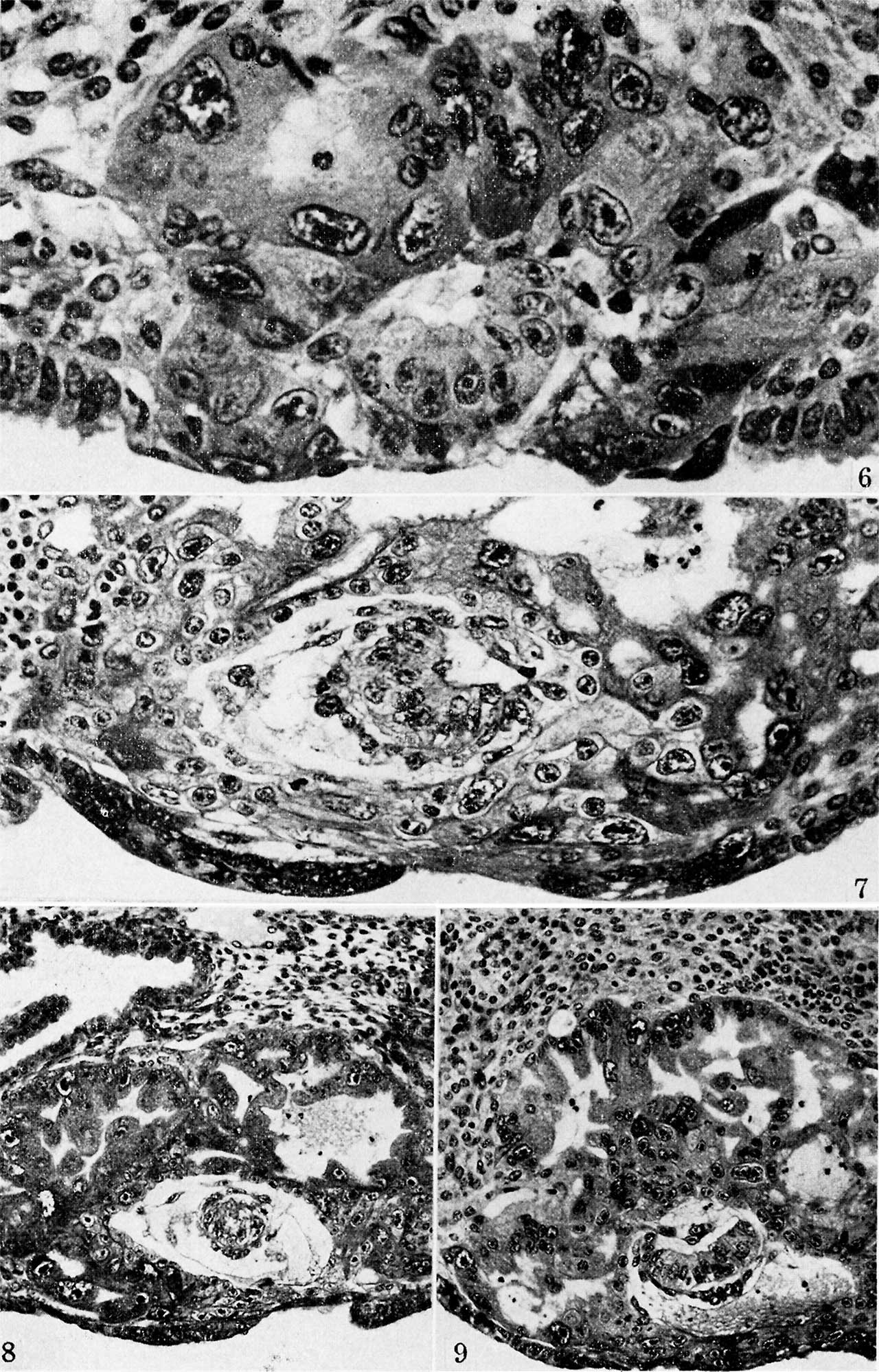
Plate 2. Ova of the ninth and tenth days
Fig. 6. A mid-crossi section from an ovum of approximately 8 days of age. Note the prominent amniotic cavity above the primitive ectoderm of the germ-disk but the amniogenic cells have not yet begun to form. There are, however, a few mesoblastic cells to the right of the germ-disk, several of which are still attached to their parent trophoblast. Carnegie 8155, section 4-4-8, X 500.
Fig. 7. A mid-cross section of an ovum 8 to 9 days of age. Note that the amniotic cavity is no more advanced than is that of the younger specimen seen in ii . 6, although there are several amniogenic cells arising from the adjacent trophoblast in the ol er specimen. Likewise, mesoblastic formation is more advanced, as shown by the presence of an early exocoelomic membrane. In places the mesoblastic cells of the latter are still attached to the trophoblast from which they appear to be originating. Carnegie 8171, section 3-2-12, X 300.
Fig. 8. A mid-cross section of an ovum of 8 to 9 days of age. The amniotic cavity is a mere slit although the amniogenic cells are well formed and have become detached from the adjacent trophoblast. The exocoelomic membrane is similar in appearance and stage of development to that of specimen shown in fig. 7. Carnegie 8215, section 12-4-5, X 140.
Fig. 9. A mid-cross section of a 9.5 -day ovum. The amniotic cavity is well formed, as are the enclosing amniogenic cells although the latter are still attached in places to their parent trophoblast. Mesoblastic formation is scanty and is represented in this section by two delaininating cel s seen toward the lower left, of the chorionic cavity. The granular material in the latter is extravasated blood which is a possible factor in delaying the formation of the mesoblast. Carnegie 8004, section 11-4-4, x 160.


Reference
Hertig AT. On the development of the amnion and exocoelomic membrane in the previllous human ovum. (1945) Yale J Biol Med. 18:107-15. PubMed 21007544
Cite this page: Hill, M.A. (2024, April 26) Embryology Hertig1945d plate02.jpg. Retrieved from https://embryology.med.unsw.edu.au/embryology/index.php/File:Hertig1945d_plate02.jpg
- © Dr Mark Hill 2024, UNSW Embryology ISBN: 978 0 7334 2609 4 - UNSW CRICOS Provider Code No. 00098G
File history
Click on a date/time to view the file as it appeared at that time.
| Date/Time | Thumbnail | Dimensions | User | Comment | |
|---|---|---|---|---|---|
| current | 13:42, 24 October 2017 | | 1,280 × 1,998 (483 KB) | Z8600021 (talk | contribs) | |
| 13:40, 24 October 2017 |  | 1,359 × 2,227 (678 KB) | Z8600021 (talk | contribs) |
You cannot overwrite this file.
File usage
The following page uses this file: