File:HansonAnson1962 fig04.jpg
Original file (1,280 × 598 pixels, file size: 201 KB, MIME type: image/jpeg)
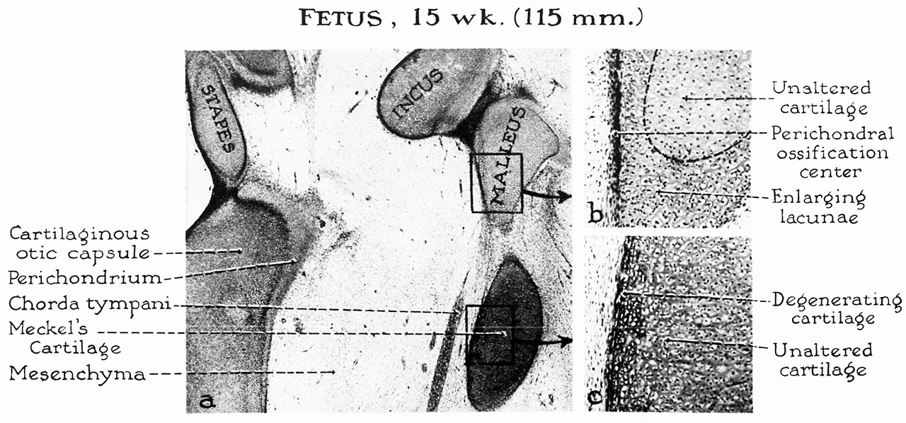
Fig. 4. Fetus 15 week 115 mm
a, In the 15-week fetus (115 mm.) the malleus begins to ossify from a single center located medially, nee the neck of the ossicles. The incus, at this level, is almost completely surrounded by a perichondral ayer.
Although the maximum growth of Meckel’s cartilage is not attained until the 17-or-18-week stage, the cartilage at the periphery of the bar shows early evidence of degeneration in the stage here illustrated.
b, Detailed examination (of the area blocked in a) reveals a layer of osteoblasts which will lay down a plaque of perichondral bone. This will spread rapidly over the ossicle. Calcification of cartilage matrix and primary resorption will ensue prior to actual endochondral ossification.
c, The cells near the periphery of Meckel’s cartilage (in the rectangular portion of a) flatten and assume an arrangement parallel to the surface of the bar. This stage in differentiation precedes their conversion into the fibrous tissue of the anterior ligament of the malleus.
Reference
Hanson JR. and Anson BJ. Development of the malleus of the human ear; Illustrated in atlas series. (1962) Q Bull Northwest Univ Med Sch. 36(2): 119–137. PMID: 13904457.
Cite this page: Hill, M.A. (2024, April 26) Embryology HansonAnson1962 fig04.jpg. Retrieved from https://embryology.med.unsw.edu.au/embryology/index.php/File:HansonAnson1962_fig04.jpg
- © Dr Mark Hill 2024, UNSW Embryology ISBN: 978 0 7334 2609 4 - UNSW CRICOS Provider Code No. 00098G
File history
Click on a date/time to view the file as it appeared at that time.
| Date/Time | Thumbnail | Dimensions | User | Comment | |
|---|---|---|---|---|---|
| current | 10:25, 7 January 2019 | | 1,280 × 598 (201 KB) | Z8600021 (talk | contribs) | |
| 10:23, 7 January 2019 |  | 1,856 × 1,253 (461 KB) | Z8600021 (talk | contribs) |
You cannot overwrite this file.
File usage
The following 2 pages use this file: