File:Hamilton1942 plate02.jpg
Original file (1,280 × 2,030 pixels, file size: 418 KB, MIME type: image/jpeg)
Plate 2
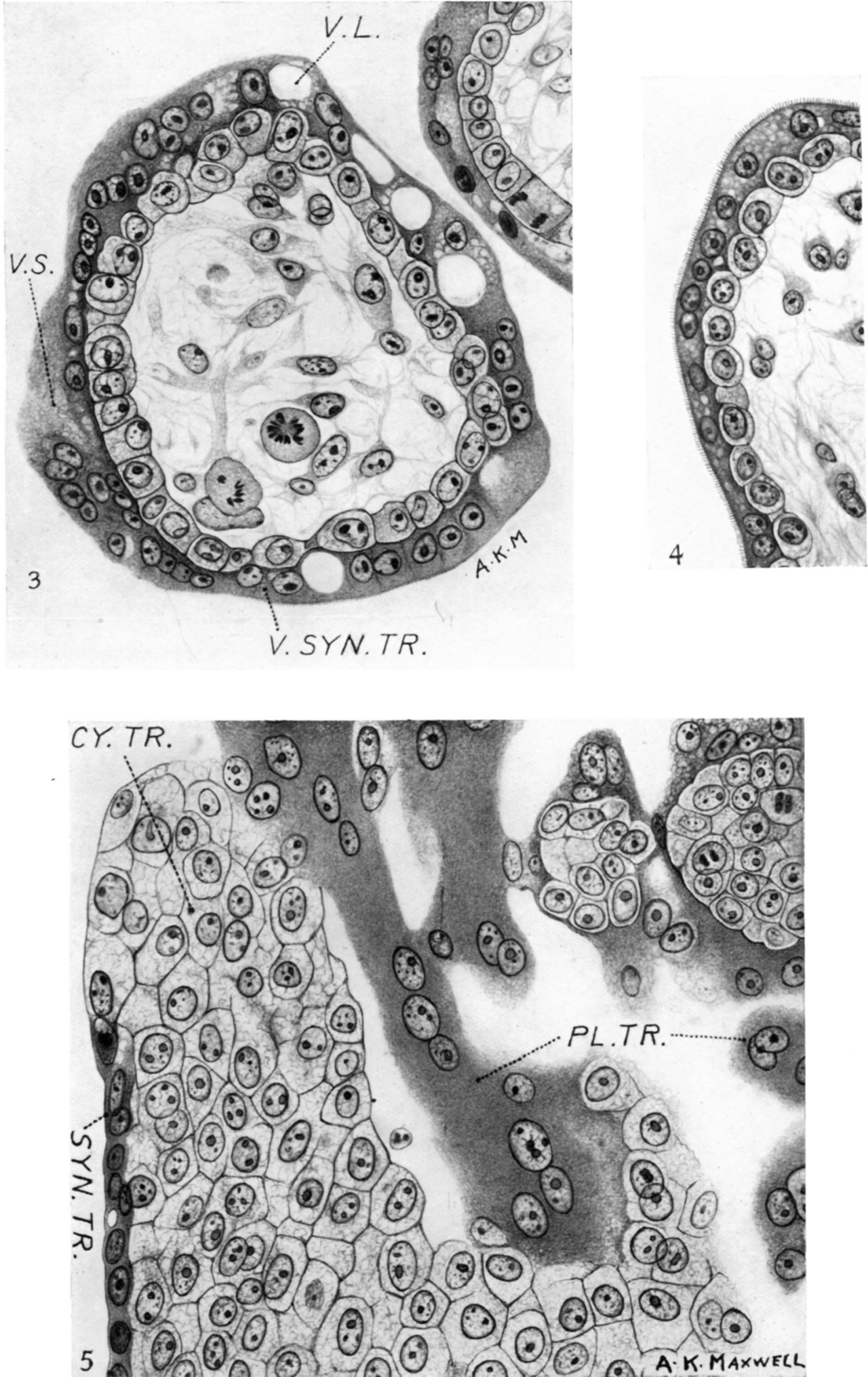
3. Transverse section through a typical villus showing the villous syncytiotrophoblast. Note large and small vacuoles and the ‘brush border’ on the bottom left-hand side of the figure. The nuclei aresmaller than those of the cytotrophohlast and tend to be disposed in pairs which are in contact or close together and are approximately of the same size. A conspicuous single nucleolus, or two true nucleoli, are present, more especially at the ends of a row of nuclei, suggesting amitotic division. x c. 550.
4. Typical ‘brush border’ on the syncytiotrophoblast of a villus. x c. 550.
5. Typical appearance of the cells of the cytotrophoblastic columns, showing bare areas and areas covered by syncytiotrophoblast. To the left and above in the figure are columns and masses which are probably remnants of the plasmoditrophoblast. x c. 550.
Reference
Hamilton WJ. and Gladstone RJ. A presomite human embryo (Shaw) - the implantation. (1942) J Anat. 76(2): 187-203 PMID 17104888
Cite this page: Hill, M.A. (2024, April 26) Embryology Hamilton1942 plate02.jpg. Retrieved from https://embryology.med.unsw.edu.au/embryology/index.php/File:Hamilton1942_plate02.jpg
- © Dr Mark Hill 2024, UNSW Embryology ISBN: 978 0 7334 2609 4 - UNSW CRICOS Provider Code No. 00098G
File history
Click on a date/time to view the file as it appeared at that time.
| Date/Time | Thumbnail | Dimensions | User | Comment | |
|---|---|---|---|---|---|
| current | 16:11, 26 February 2017 | | 1,280 × 2,030 (418 KB) | Z8600021 (talk | contribs) | |
| 16:10, 26 February 2017 |  | 1,574 × 2,546 (1.12 MB) | Z8600021 (talk | contribs) |
You cannot overwrite this file.
File usage
The following 2 pages use this file: