File:Gray1113.jpg
Gray1113.jpg (600 × 385 pixels, file size: 68 KB, MIME type: image/jpeg)
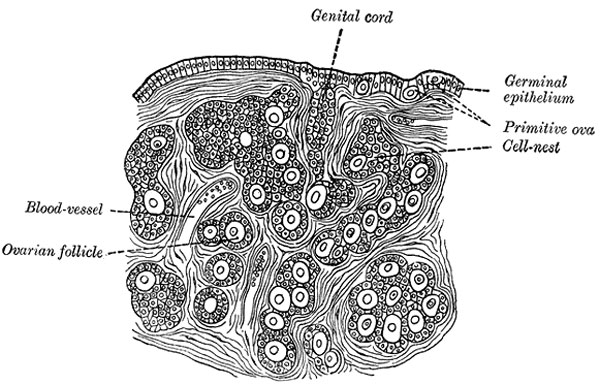
Section of the Ovary of a Newly Born Child
(Waldeyer.)
| Historic Disclaimer - information about historic embryology pages |
|---|
 |
--Mark Hill 08:54, 28 Feb 2010 (EST) Note this historic drawing wrongly ascribes the "germinal epithelium" as the source of oocytes and follicles.The surface of the ovary is covered by a single layer of cuboidal epithelium (the germinal epithelium) that is continuous with the peritoneal mesothelium. Fibrous connective tissue forms a thin capsule, the tunica albuginea, immediately beneath the epithelium.
- Gray's Images: Development | Lymphatic | Neural | Vision | Hearing | Somatosensory | Integumentary | Respiratory | Gastrointestinal | Urogenital | Endocrine | Surface Anatomy | iBook | Historic Disclaimer
| Historic Disclaimer - information about historic embryology pages |
|---|
|
| iBook - Gray's Embryology | |
|---|---|

|
|
Reference
Gray H. Anatomy of the human body. (1918) Philadelphia: Lea & Febiger.
Cite this page: Hill, M.A. (2024, April 26) Embryology Gray1113.jpg. Retrieved from https://embryology.med.unsw.edu.au/embryology/index.php/File:Gray1113.jpg
- © Dr Mark Hill 2024, UNSW Embryology ISBN: 978 0 7334 2609 4 - UNSW CRICOS Provider Code No. 00098G
File history
Click on a date/time to view the file as it appeared at that time.
| Date/Time | Thumbnail | Dimensions | User | Comment | |
|---|---|---|---|---|---|
| current | 08:51, 28 May 2011 | | 600 × 385 (68 KB) | S8600021 (talk | contribs) | ==Section of the Ovary of a Newly Born Child== (Waldeyer.) {{Historic Disclaimer}} (text modified from Gray's Anatomy) {{Gray Anatomy}} Category:Human Category:Genital Category:Female Category:Ovary Category:Neonatal |
You cannot overwrite this file.
File usage
The following 6 pages use this file: