File:Gray1109.jpg
From Embryology
Gray1109.jpg (464 × 487 pixels, file size: 56 KB, MIME type: image/jpeg)
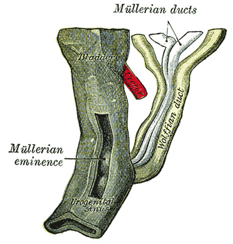
Fig. 1109. Urogenital Sinus of Female Human Embryo of 8.5 to 9 weeks old
(From model by Keibel)
| Historic Disclaimer - information about historic embryology pages |
|---|
| Pages where the terms "Historic" (textbooks, papers, people, recommendations) appear on this site, and sections within pages where this disclaimer appears, indicate that the content and scientific understanding are specific to the time of publication. This means that while some scientific descriptions are still accurate, the terminology and interpretation of the developmental mechanisms reflect the understanding at the time of original publication and those of the preceding periods, these terms, interpretations and recommendations may not reflect our current scientific understanding. (More? Embryology History | Historic Embryology Papers) |
The Müllerian Ducts (Paramesonephric Ducts)
- Shortly after the formation of the Wolffian ducts a second pair of ducts is developed, the Müllerian ducts
- Each arises on the lateral aspect of the corresponding Wolffian duct as a tubular invagination of the cells lining the coelom
- orifice of the invagination remains patent, and undergoes enlargement and modification to form the abdominal ostium of the uterine tube
- ducts pass backward lateral to the Wolffian ducts
- toward the posterior end of the embryo they cross to the medial side of these ducts
- come to lie side by side between and behind the latter
- the four ducts forming what is termed the genital cord
- Müllerian ducts end in an epithelial elevation, the Müllerian eminence
- on the ventral part of the cloaca between the orifices of the Wolffian duct
- at a later date they open into the cloaca in this situation
(Text modified from Gray's Anatomy)
- Links: uterus | Gray's Urogenital Images
- Gray's Images: Development | Lymphatic | Neural | Vision | Hearing | Somatosensory | Integumentary | Respiratory | Gastrointestinal | Urogenital | Endocrine | Surface Anatomy | iBook | Historic Disclaimer
| Historic Disclaimer - information about historic embryology pages |
|---|
| Pages where the terms "Historic" (textbooks, papers, people, recommendations) appear on this site, and sections within pages where this disclaimer appears, indicate that the content and scientific understanding are specific to the time of publication. This means that while some scientific descriptions are still accurate, the terminology and interpretation of the developmental mechanisms reflect the understanding at the time of original publication and those of the preceding periods, these terms, interpretations and recommendations may not reflect our current scientific understanding. (More? Embryology History | Historic Embryology Papers) |
| iBook - Gray's Embryology | |
|---|---|

|
|
Reference
Gray H. Anatomy of the human body. (1918) Philadelphia: Lea & Febiger.
Cite this page: Hill, M.A. (2024, April 26) Embryology Gray1109.jpg. Retrieved from https://embryology.med.unsw.edu.au/embryology/index.php/File:Gray1109.jpg
- © Dr Mark Hill 2024, UNSW Embryology ISBN: 978 0 7334 2609 4 - UNSW CRICOS Provider Code No. 00098G
File history
Click on a date/time to view the file as it appeared at that time.
| Date/Time | Thumbnail | Dimensions | User | Comment | |
|---|---|---|---|---|---|
| current | 09:27, 28 May 2011 |  | 464 × 487 (56 KB) | S8600021 (talk | contribs) | ==Urogenital Sinus of Female Human Embryo of 8.5 to 9 weeks old== (From model by Keibel) {{Gray Anatomy}} Category:Human Category:Genital Category:Female Category:Uterus |
You cannot overwrite this file.
File usage
The following 6 pages use this file:

