File:Gray1027.jpg
Gray1027.jpg (598 × 600 pixels, file size: 101 KB, MIME type: image/jpeg)
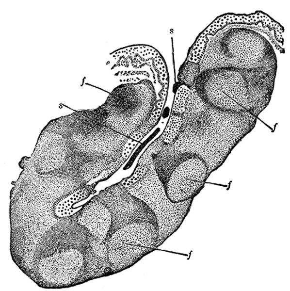
Section through one of the crypts of the tonsil
(Stöhr.) Magnified. e. Stratified epithelium of general surface, continued into crypt. f, f.
Nodules of lymphoid tissue—opposite each nodule numbers of lymph cells are passing into or through the epithelium. s, s. Cells which have thus escaped to mix with the saliva as salivary corpuscles.
Structure
The follicles of the tonsil are lined by a continuation of the mucous membrane of the pharynx, covered with stratified squamous epithelium; around each follicle is a layer of closed capsules consisting of lymphoid tissue imbedded in the submucous tissue. Lymph corpuscles are found in large numbers invading the stratified epithelium. It is probable that they pass into the mouth and form the so-called salivary corpusles. Surrounding each follicle is a close plexus of lymphatics, from which the lymphatic vessels pass to the deep cervical glands in the neighborhood of the greater cornu of the hyoid bone, behind and below the angle of the mandible.
- Gray's Images: Development | Lymphatic | Neural | Vision | Hearing | Somatosensory | Integumentary | Respiratory | Gastrointestinal | Urogenital | Endocrine | Surface Anatomy | iBook | Historic Disclaimer
| Historic Disclaimer - information about historic embryology pages |
|---|
| Pages where the terms "Historic" (textbooks, papers, people, recommendations) appear on this site, and sections within pages where this disclaimer appears, indicate that the content and scientific understanding are specific to the time of publication. This means that while some scientific descriptions are still accurate, the terminology and interpretation of the developmental mechanisms reflect the understanding at the time of original publication and those of the preceding periods, these terms, interpretations and recommendations may not reflect our current scientific understanding. (More? Embryology History | Historic Embryology Papers) |
| iBook - Gray's Embryology | |
|---|---|

|
|
Reference
Gray H. Anatomy of the human body. (1918) Philadelphia: Lea & Febiger.
Cite this page: Hill, M.A. (2024, April 27) Embryology Gray1027.jpg. Retrieved from https://embryology.med.unsw.edu.au/embryology/index.php/File:Gray1027.jpg
- © Dr Mark Hill 2024, UNSW Embryology ISBN: 978 0 7334 2609 4 - UNSW CRICOS Provider Code No. 00098G
File history
Click on a date/time to view the file as it appeared at that time.
| Date/Time | Thumbnail | Dimensions | User | Comment | |
|---|---|---|---|---|---|
| current | 13:46, 25 February 2013 |  | 598 × 600 (101 KB) | Z8600021 (talk | contribs) | ==Section through one of the crypts of the tonsil== (Stöhr.) Magnified. e. Stratified epithelium of general surface, continued into crypt. f, f. Nodules of lymphoid tissue—opposite each nodule numbers of lymph cells are passing into or through the |
You cannot overwrite this file.
File usage
The following page uses this file:

