File:Gray0942.jpg
Gray0942.jpg (800 × 515 pixels, file size: 127 KB, MIME type: image/jpeg)
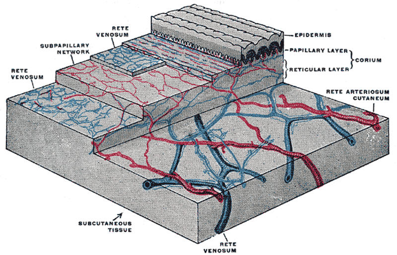
The distribution of the bloodvessels in the skin of the sole of the foot
(Spalteholz.)
Reticular layer
(stratum reticulare; deep layer) consists of strong interlacing bands, composed chiefly of white fibrous tissue, but containing some fibers of yellow elastic tissue, which vary in number in different parts; and connective-tissue corpuscles, which are often to be found flattened against the white fibrous tissue bundles. Toward the attached surface the fasciculi are large and coarse, and the areolæ left by their interlacement are large, and occupied by adipose tissue and sweat glands. Below the reticular layer is the subcutaneous areolar tissue, which, except in a few situations, contains fat.
Papillary layer
(stratum papillare; superficial layer; corpus papillare of the corium) consists of numerous small, highly sensitive, and vascular eminences, the papillæ, which rise perpendicularly from its surface. The papillæ are minute conical eminences, having rounded or blunted extremities, occasionally divided into two or more parts, and are received into corresponding pits on the under surface of the cuticle. On the general surface of the body, more especially in parts endowed with slight sensibility, they are few in number, and exceedingly minute; but in some situations, as upon the palmar surfaces of the hands and fingers, and upon the plantar surfaces of the feet and toes, they are long, of large size, closely aggregated together, and arranged in parallel curved lines, forming the elevated ridges seen on the free surface of the epidermis. Each ridge contains two rows of papillæ, between which the ducts of the sudoriferous glands pass outward to open on the summit of the ridge. Each papilla consists of very small and closely interlacing bundles of finely fibrillated tissue, with a few elastic fibers; within this tissue is a capillary loop, and in some papillæ, especially in the palms of the hands and the fingers, there are tactile corpuscles. (Text modified from Gray's 1918 Anatomy)
- Gray's Images: Development | Lymphatic | Neural | Vision | Hearing | Somatosensory | Integumentary | Respiratory | Gastrointestinal | Urogenital | Endocrine | Surface Anatomy | iBook | Historic Disclaimer
| Historic Disclaimer - information about historic embryology pages |
|---|
 |
| iBook - Gray's Embryology | |
|---|---|

|
|
Reference
Gray H. Anatomy of the human body. (1918) Philadelphia: Lea & Febiger.
Cite this page: Hill, M.A. (2024, April 27) Embryology Gray0942.jpg. Retrieved from https://embryology.med.unsw.edu.au/embryology/index.php/File:Gray0942.jpg
- © Dr Mark Hill 2024, UNSW Embryology ISBN: 978 0 7334 2609 4 - UNSW CRICOS Provider Code No. 00098G
File history
Click on a date/time to view the file as it appeared at that time.
| Date/Time | Thumbnail | Dimensions | User | Comment | |
|---|---|---|---|---|---|
| current | 23:41, 19 August 2012 | | 800 × 515 (127 KB) | Z8600021 (talk | contribs) | ==The distribution of the bloodvessels in the skin of the sole of the foot== (Spalteholz.) (Text modified from Gray's 1918 Anatomy) {{Gray Anatomy}} Category:Cartoon Category:Senses Category:Somatosensory Category:Human [[Category:Int |
You cannot overwrite this file.
File usage
The following page uses this file: