File:Gray0890.jpg

Original file (700 × 700 pixels, file size: 104 KB, MIME type: image/jpeg)
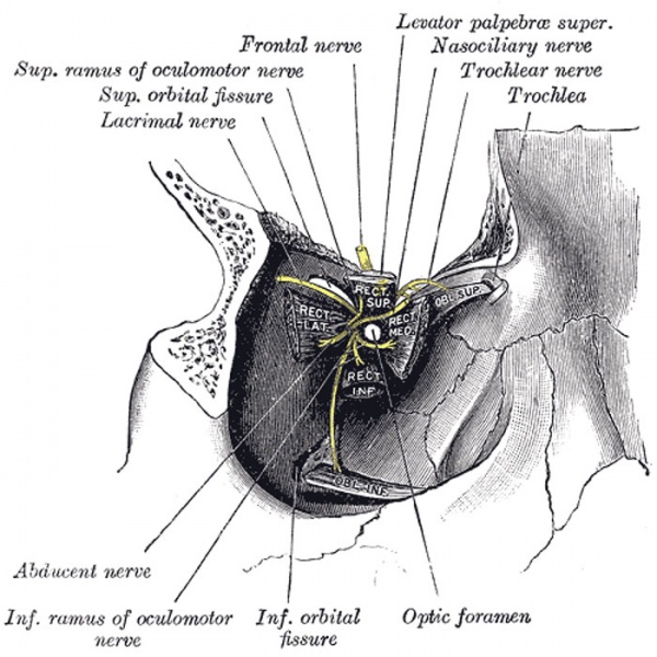
Right Ocular Muscles
Dissection showing origins of right ocular muscles, and nerves entering by the superior orbital fissure.
The four Recti (Fig. 889) arise from a fibrous ring (annulus tendineus communis) which surrounds the upper, medial, and lower margins of the optic foramen and encircles the optic nerve (Fig. 890). The ring is completed by a tendinous bridge prolonged over the lower and medial part of the superior orbital fissure and attached to a tubercle on the margin of the great wing of the sphenoid, bounding the fissure. Two specialized parts of this fibrous ring may be made out: a lower, the ligament or tendon of Zinn, which gives origin to the Rectus inferior, part of the Rectus internus, and the lower head of origin of the Rectus lateralis; and an upper, which gives origin to the Rectus superior, the rest of the Rectus medialis, and the upper head of the Rectus lateralis. This upper band is sometimes termed the superior tendon of Lockwood. Each muscle passes forward in the position implied by its name, to be inserted by a tendinous expansion into the sclera, about 6 mm. from the margin of the cornea. Between the two heads of the Rectus lateralis is a narrow interval, through which pass the two divisions of the oculomotor nerve, the nasociliary nerve, the abducent nerve, and the ophthalmic vein. Although these muscles present a common origin and are inserted in a similar manner into the sclera, there are certain differences to be observed in them as regards their length and breadth. The Rectus medialis is the broadest, the Rectus lateralis the longest, and the Rectus superior the thinnest and narrowest.
(Text modified from Gray's 1918 Anatomy)
- Gray's Images: Development | Lymphatic | Neural | Vision | Hearing | Somatosensory | Integumentary | Respiratory | Gastrointestinal | Urogenital | Endocrine | Surface Anatomy | iBook | Historic Disclaimer
| Historic Disclaimer - information about historic embryology pages |
|---|
 |
| iBook - Gray's Embryology | |
|---|---|

|
|
Reference
Gray H. Anatomy of the human body. (1918) Philadelphia: Lea & Febiger.
Cite this page: Hill, M.A. (2024, April 26) Embryology Gray0890.jpg. Retrieved from https://embryology.med.unsw.edu.au/embryology/index.php/File:Gray0890.jpg
- © Dr Mark Hill 2024, UNSW Embryology ISBN: 978 0 7334 2609 4 - UNSW CRICOS Provider Code No. 00098G
File history
Click on a date/time to view the file as it appeared at that time.
| Date/Time | Thumbnail | Dimensions | User | Comment | |
|---|---|---|---|---|---|
| current | 22:43, 19 August 2012 | | 700 × 700 (104 KB) | Z8600021 (talk | contribs) | ==Right Ocular Muscles== Dissection showing origins of right ocular muscles, and nerves entering by the superior orbital fissure. (Text modified from Gray's 1918 Anatomy) {{Gray Anatomy}} Category:Cartoon Category:Senses Category:Vision |
You cannot overwrite this file.
File usage
The following page uses this file: