File:Gray0882.jpg
Original file (800 × 649 pixels, file size: 123 KB, MIME type: image/jpeg)
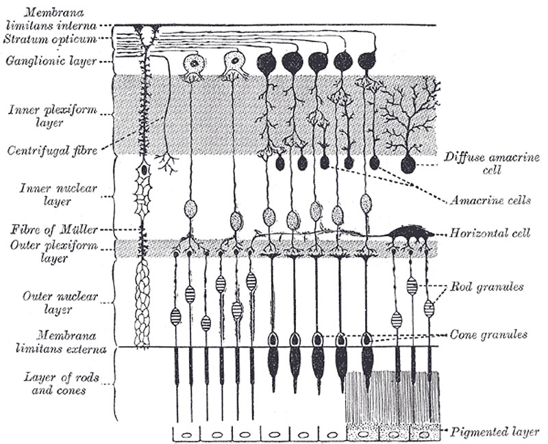
Plan of Retinal Neurons
(After Cajal.)
1. The stratum opticum or layer of nerve fibers is formed by the expansion of the fibers of the optic nerve; it is thickest near the porus opticus, gradually diminishing toward the ora serrata. As the nerve fibers pass through the lamina cribrosa scleræ they lose their medullary sheaths and are continued onward through the choroid and retina as simple axis-cylinders. When they reach the internal surface of the retina they radiate from their point of entrance over this surface grouped in bundles, and in many places arranged in plexuses. Most of the fibers are centripetal, and are the direct continuations of the axis-cylinder processes of the cells of the ganglionic layer, but a few of them are centrifugal and ramify in the inner plexiform and inner nuclear layers, where they end in enlarged extremities.
2. The ganglionic layer consists of a single layer of large ganglion cells, except in the macula lutea, where there are several strata. The cells are somewhat flask-shaped; the rounded internal surface of each resting on the stratum opticum, and sending off an axon which is prolonged into it. From the opposite end numerous dendrites extend into the inner plexiform layer, where they branch and form flattened arborizations at different levels. The ganglion cells vary much in size, and the dendrites of the smaller ones as a rule arborize in the inner plexiform layer as soon as they enter it; while those of the larger cells ramify close to the inner nuclear layer.
3. The inner plexiform layer is made up of a dense reticulum of minute fibrils formed by the interlacement of the dendrites of the ganglion cells with those of the cells of the inner nuclear layer; within this reticulum a few branched spongioblasts are sometimes imbedded.
4. The inner nuclear layer or layer of inner granules is made up of a number of closely packed cells, of which there are three varieties, viz.: bipolar cells, horizontal cells, and amacrine cells.
The bipolar cells, by far the most numerous, are round or oval in shape, and each is prolonged into an inner and an outer process. They are divisible into rod bipolars and cone bipolars. The inner processes of the rod bipolars run through the inner plexiform layer and arborize around the bodies of the cells of the ganglionic layer; their outer processes end in the outer plexiform layer in tufts of fibrils around the button-like ends of the inner processes of the rod granules. The inner processes of the cone bipolars ramify in the inner plexiform layer in contact with the dendrites of the ganglionic cells.
The horizontal cells lie in the outer part of the inner nuclear layer and possess somewhat flattened cell bodies. Their dendrites divide into numerous branches in the outer plexiform layer, while their axons run horizontally for some distance and finally ramify in the same layer.
The amacrine cells are placed in the inner part of the inner nuclear layer, and are so named because they have not yet been shown to possess axis-cylinder processes. Their dendrites undergo extensive ramification in the inner plexiform layer.
5. The outer plexiform layer is much thinner than the inner; but, like it, consists of a dense net-work of minute fibrils derived from the processes of the horizontal cells of the preceding layer, and the outer processes of the rod and cone bipolar granules, which ramify in it, forming arborizations around the enlarged ends of the rod fibers and with the branched foot plates of the cone fibers.
6. The outer nuclear layer or layer of outer granules, like the inner nuclear layer, contains several strata of oval nuclear bodies; they are of two kinds, viz.: rod and cone granules, so named on account of their being respectively connected with the rods and cones of the next layer. The rod granules are much the more numerous, and are placed at different levels throughout the layer. Their nuclei present a peculiar cross-striped appearance, and prolonged from either extremity of each cell is a fine process; the outer process is continuous with a single rod of the layer of rods and cones; the inner ends in the outer plexiform layer in an enlarged extremity, and is imbedded in the tuft into which the outer processes of the rod bipolar cells break up. In its course it presents numerous varicosities. The cone granules, fewer in number than the rod granules, are placed close to the membrana limitans externa, through which they are continuous with the cones of the layer of rods and cones. They do not present any cross-striation, but contain a pyriform nucleus, which almost completely fills the cell. From the inner extremity of the granule a thick process passes into the outer plexiform layer, and there expands into a pyramidal enlargement or foot plate, from which are given off numerous fine fibrils, that come in contact with the outer processes of the cone bipolars.
7. The Layer of Rods and Cones (Jacob’s membrane).—The elements composing this layer are of two kinds, rods and cones, the former being much more numerous than the latter except in the macula lutea. The rods are cylindrical, of nearly uniform thickness, and are arranged perpendicularly to the surface. Each rod consists of two segments, an outer and inner, of about equal lengths. The segments differ from each other as regards refraction and in their behavior toward coloring reagents; the inner segment is stained by carmine, iodine, etc.; the outer segment is not stained by these reagents, but is colored yellowish brown by osmic acid. The outer segment is marked by transverse striæ, and tends to break up into a number of thin disks superimposed on one another; it also exhibits faint longitudinal markings. The deeper part of the inner segment is indistinctly granular; its more superficial part presents a longitudinal striation, being composed of fine, bright, highly refracting fibrils. The visual purple or rhodopsin is found only in the outer segments.
The cones are conical or flask-shaped, their broad ends resting upon the membrana limitans externa, the narrow-pointed extremity being turned to the choroid. Like the rods, each is made up of two segments, outer and inner; the outer segment is a short conical process, which, like the outer segment of the rod, exhibits transverse striæ. The inner segment resembles the inner segment of the rods in structure, presenting a superficial striated and deep granular part, but differs from it in size and shape, being bulged out laterally and flask-shaped. The chemical and optical characters of the two portions are identical with those of the rods.
(Text modified from Gray's 1918 Anatomy)
- Gray's Images: Development | Lymphatic | Neural | Vision | Hearing | Somatosensory | Integumentary | Respiratory | Gastrointestinal | Urogenital | Endocrine | Surface Anatomy | iBook | Historic Disclaimer
| Historic Disclaimer - information about historic embryology pages |
|---|
 |
| iBook - Gray's Embryology | |
|---|---|

|
|
Reference
Gray H. Anatomy of the human body. (1918) Philadelphia: Lea & Febiger.
Cite this page: Hill, M.A. (2024, April 27) Embryology Gray0882.jpg. Retrieved from https://embryology.med.unsw.edu.au/embryology/index.php/File:Gray0882.jpg
- © Dr Mark Hill 2024, UNSW Embryology ISBN: 978 0 7334 2609 4 - UNSW CRICOS Provider Code No. 00098G
File history
Click on a date/time to view the file as it appeared at that time.
| Date/Time | Thumbnail | Dimensions | User | Comment | |
|---|---|---|---|---|---|
| current | 15:05, 19 August 2012 | | 800 × 649 (123 KB) | Z8600021 (talk | contribs) | 1. The stratum opticum or layer of nerve fibers is formed by the expansion of the fibers of the optic nerve; it is thickest near the porus opticus, gradually diminishing toward the ora serrata. As the nerve fibers pass through the lamina cribrosa scleræ |
You cannot overwrite this file.
File usage
The following page uses this file: