File:Gray0877.jpg

Original file (500 × 794 pixels, file size: 101 KB, MIME type: image/jpeg)
Blood Vessels of the Eye
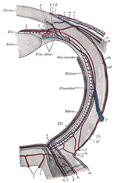
Diagram of the blood vessels of the eye, as seen in a horizontal section. (Leber, after Stöhr.).
Course of vasa centralia retinæ: a. Arteria. a. 147 Vena centralis retinæ. B. Anastomosis with vessels of outer coats. C. Anastomosis with branches of short posterior ciliary arteries. D. Anastomosis with chorioideal vessels. Course of vasa ciliar. postic. brev.: I. Arteriæ, and I1. Venæ ciliar. postic. brev. II. Episcleral artery. II1. Episcleral vein. III. Capillaries of lamina choriocapillaris. Course of vasa ciliar. postic. long.: 1. a. ciliar. post. longa. 2. Circulus iridis major cut across. 3. Branches to ciliary body. 4. Branches to iris. Course of vasa ciliar. ant.: a. Arteria. a1. Vena ciliar. ant. b. Junction with the circulus iridis major. c. Junction with lamina choriocapill. d. Arterial, and d1. Venous episcleral branches. e. Arterial, and e1. Venous branches to conjunctiva scleræ. f. Arterial, and f1. Venous branches to corneal border. V. Vena vorticosa. S. Transverse section of sinus venosus scleræ.
(Text modified from Gray's 1918 Anatomy)
- Gray's Images: Development | Lymphatic | Neural | Vision | Hearing | Somatosensory | Integumentary | Respiratory | Gastrointestinal | Urogenital | Endocrine | Surface Anatomy | iBook | Historic Disclaimer
| Historic Disclaimer - information about historic embryology pages |
|---|
| Pages where the terms "Historic" (textbooks, papers, people, recommendations) appear on this site, and sections within pages where this disclaimer appears, indicate that the content and scientific understanding are specific to the time of publication. This means that while some scientific descriptions are still accurate, the terminology and interpretation of the developmental mechanisms reflect the understanding at the time of original publication and those of the preceding periods, these terms, interpretations and recommendations may not reflect our current scientific understanding. (More? Embryology History | Historic Embryology Papers) |
| iBook - Gray's Embryology | |
|---|---|

|
|
Reference
Gray H. Anatomy of the human body. (1918) Philadelphia: Lea & Febiger.
Cite this page: Hill, M.A. (2024, April 26) Embryology Gray0877.jpg. Retrieved from https://embryology.med.unsw.edu.au/embryology/index.php/File:Gray0877.jpg
- © Dr Mark Hill 2024, UNSW Embryology ISBN: 978 0 7334 2609 4 - UNSW CRICOS Provider Code No. 00098G
File history
Click on a date/time to view the file as it appeared at that time.
| Date/Time | Thumbnail | Dimensions | User | Comment | |
|---|---|---|---|---|---|
| current | 09:38, 19 August 2012 |  | 500 × 794 (101 KB) | Z8600021 (talk | contribs) | ==Blood Vessels of the Eye== Diagram of the blood vessels of the eye, as seen in a horizontal section. (Leber, after Stöhr.). Course of vasa centralia retinæ: a. Arteria. a. 147 Vena centralis retinæ. B. Anastomosis with vessels of outer coats. C. An |
You cannot overwrite this file.
File usage
The following page uses this file:
