File:GladstoneHamilton1941 text-fig03.jpg
From Embryology

Size of this preview: 739 × 600 pixels. Other resolution: 1,280 × 1,039 pixels.
Original file (1,280 × 1,039 pixels, file size: 212 KB, MIME type: image/jpeg)
Text-fig. 3. Idealized reconstruction of a median sagittal section of the embryo
Showing the right half of the embryonic rudiment and the attachment of the connecting stalk. magnification of 100 diameters
Reference
Gladstone RJ. and Hamilton WJ. A presomite human embryo (Shaw) with primitive streak and chorda canal with special reference to the development of the vascular system. (1941) Amer. J Anat. 76(1): 9-44.
Cite this page: Hill, M.A. (2024, April 26) Embryology GladstoneHamilton1941 text-fig03.jpg. Retrieved from https://embryology.med.unsw.edu.au/embryology/index.php/File:GladstoneHamilton1941_text-fig03.jpg
- © Dr Mark Hill 2024, UNSW Embryology ISBN: 978 0 7334 2609 4 - UNSW CRICOS Provider Code No. 00098G
File history
Click on a date/time to view the file as it appeared at that time.
| Date/Time | Thumbnail | Dimensions | User | Comment | |
|---|---|---|---|---|---|
| current | 16:42, 26 February 2017 | | 1,280 × 1,039 (212 KB) | Z8600021 (talk | contribs) | |
| 16:40, 26 February 2017 | 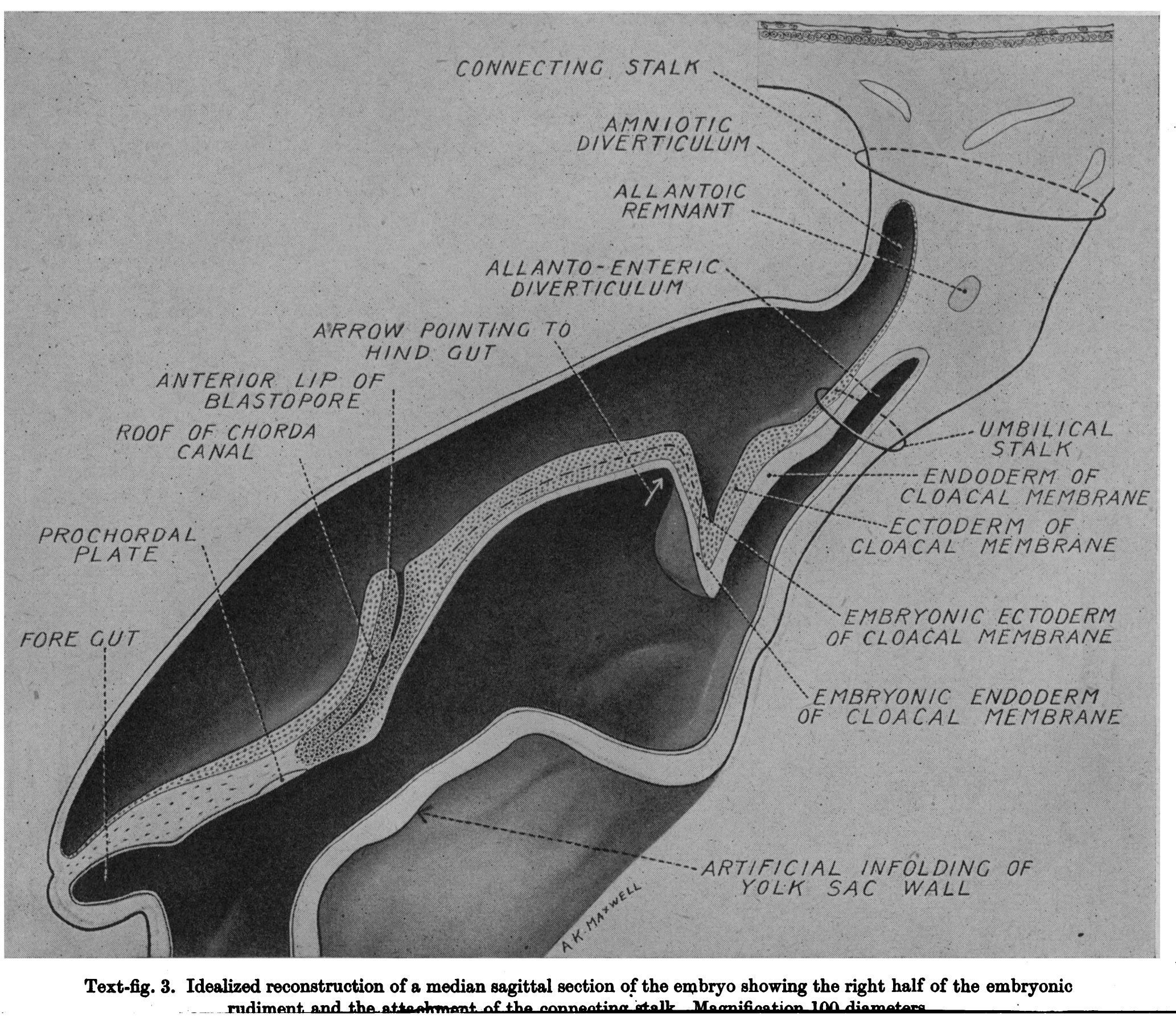 | 2,066 × 1,808 (1.01 MB) | Z8600021 (talk | contribs) |
You cannot overwrite this file.