File:Frazer1919 fig01.jpg
From Embryology
Size of this preview: 800 × 475 pixels. Other resolution: 1,280 × 760 pixels.
Original file (1,280 × 760 pixels, file size: 123 KB, MIME type: image/jpeg)
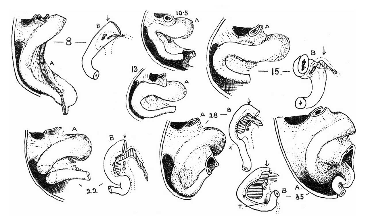
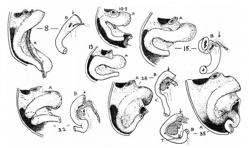
Fig. 1. The duodenum throughout the “first stage” of intestinal development
A, side view, B, front view. The numerals give the size of embryo in millimetres. See text.
| Historic Disclaimer - information about historic embryology pages |
|---|
 |
- Links: intestine
Reference
Frazer JE. The formation of the duodenal curve. J Anat. 1919 53(4):292-7. PMID 17103870
Cite this page: Hill, M.A. (2024, April 27) Embryology Frazer1919 fig01.jpg. Retrieved from https://embryology.med.unsw.edu.au/embryology/index.php/File:Frazer1919_fig01.jpg
- © Dr Mark Hill 2024, UNSW Embryology ISBN: 978 0 7334 2609 4 - UNSW CRICOS Provider Code No. 00098G
File history
Click on a date/time to view the file as it appeared at that time.
| Date/Time | Thumbnail | Dimensions | User | Comment | |
|---|---|---|---|---|---|
| current | 13:07, 15 June 2018 | | 1,280 × 760 (123 KB) | Z8600021 (talk | contribs) | |
| 13:06, 15 June 2018 | 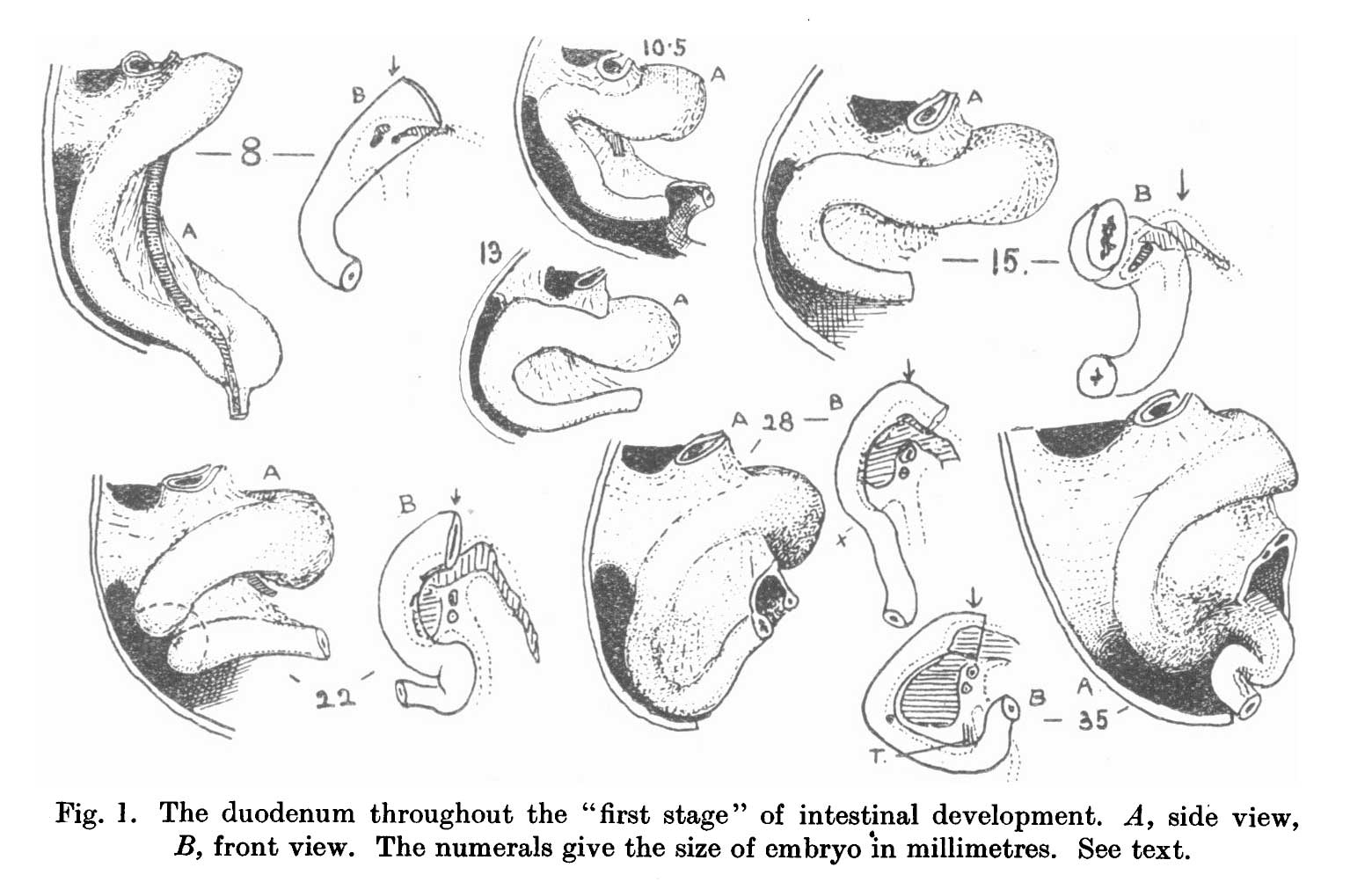 | 1,560 × 1,003 (137 KB) | Z8600021 (talk | contribs) | ===Reference=== {{Ref-Frazer1919}} {{Footer}} |
You cannot overwrite this file.
File usage
The following page uses this file: