File:Frazer1915 fig14.jpg

Original file (1,000 × 549 pixels, file size: 122 KB, MIME type: image/jpeg)
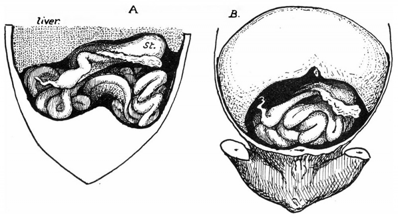
Fig. 14. Two specimens of 45 mm
Intestines exposed by raising the liver carefully and depressing the pelvic parts: in A the liver is represented as divided transversely, and the drawing is somewhat diagrammatic. In A the ca:cum lay as shown in a curved state on the coils a. little to the right of the middle, but in B it was more to the right, though still delinitely resting on the coils. St. is the stomach. The inesocolon of the loop is seen in A but not in B. The overhanging projecting part of the omental bursa has been raised to show the coils, but the left transverse colon is too deep to be shown in this way. Altogetlier live specimens of this period were examined. Of the others, one ofubout 40 mm. was like A in its caecal relations, another of 42 mm. resembled B, but in one of 39 mm. the czecuin lay to the right of the coils. in this case the whole mass of coils seemed to he carried more than is usual to the left, so that the caecum was not much beyond the level of the duodenum although it lay to the right of them, and the right lobe of the liver was perhaps larger than usual.
| Historic Disclaimer - information about historic embryology pages |
|---|
| Pages where the terms "Historic" (textbooks, papers, people, recommendations) appear on this site, and sections within pages where this disclaimer appears, indicate that the content and scientific understanding are specific to the time of publication. This means that while some scientific descriptions are still accurate, the terminology and interpretation of the developmental mechanisms reflect the understanding at the time of original publication and those of the preceding periods, these terms, interpretations and recommendations may not reflect our current scientific understanding. (More? Embryology History | Historic Embryology Papers) |
- Links: Fig 1 | Fig 2 | Fig 3 | Fig 4 | Fig 5 | Fig 6 | Fig 7 | Fig 8 | Fig 9 | Fig 10 | Fig 11 | Fig 12 | Fig 13 | Fig 14 | Fig 15 | Fig 16 | Fig 17 | Fig 18 | 1915 Frazer | Intestine Development | Category:Intestine
Reference
Frazer JE. and Robbins RH. On the factors concerned in causing rotation of the intestine in man. (1915) J Anat. 50(1): 75-110. PMID 17233053
Cite this page: Hill, M.A. (2024, April 26) Embryology Frazer1915 fig14.jpg. Retrieved from https://embryology.med.unsw.edu.au/embryology/index.php/File:Frazer1915_fig14.jpg
- © Dr Mark Hill 2024, UNSW Embryology ISBN: 978 0 7334 2609 4 - UNSW CRICOS Provider Code No. 00098G
File history
Click on a date/time to view the file as it appeared at that time.
| Date/Time | Thumbnail | Dimensions | User | Comment | |
|---|---|---|---|---|---|
| current | 05:19, 9 January 2017 |  | 1,000 × 549 (122 KB) | Z8600021 (talk | contribs) | |
| 05:11, 9 January 2017 |  | 1,454 × 1,153 (354 KB) | Z8600021 (talk | contribs) | {{Historic Disclaimer}} ===Reference=== {{Ref-Frazer1915}} {{Footer}} |
You cannot overwrite this file.
File usage
The following 4 pages use this file:
