File:Foster038.jpg
Foster038.jpg (432 × 416 pixels, file size: 46 KB, MIME type: image/jpeg)
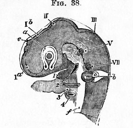
Fig. 38. Head Of A Chick Of The Third Day
Viewed Sideways As A Transparent Object. (From Huxley.)
I a. the vesicle of the cerebral hemisphere. 1 6. the vesicle of the third ventricle (the original fore-brain) ; at its summit is seen the projection of the pineal gland e.
Below this portion of the brain is seen, in optical section, the optic vesicle a already involuted with its thick inner and thinner outer wall (the letter a is placed on the junction of the two, the primary cavity being almost obliterated). In the centre of the vesicle lies the lens, the shaded portion being the expression of its cavity. Below the lens between the two limbs of the horseshoe is the choroidal fissure.
II. the mid-brain. III. the hind-brain. V. the rudiments of the fifth cranial nerve, VII. of the seventh. Below the seventh nerve is seen the auditory vesicle b. The head having been subjected to pressure, the vesicle appears somewhat distorted as if squeezed out of place. The orifice is not yet quite closed up.
I, the inferior maxillary process of the first visceral or mandibular fold. Below, and to the right of this, is seen the first visceral cleft, below that again the second visceral fold (2), and lower down the third (3) and fourth (4) visceral folds. In front of the folds (i.e. to the left) is seen the arterial end of the heart r the aortic arches being buried in their respective visceral folds.
f. represents the mesoblast of the base of the brain and spinal cord.
| Historic Disclaimer - information about historic embryology pages |
|---|
 |
Reference
Foster, M., Balfour, F. M., Sedgwick, A., & Heape, W. (1883). The Elements of Embryology. (2nd ed.). London: Macmillan and Co.
The Elements of Embryology (1883)
File history
Click on a date/time to view the file as it appeared at that time.
| Date/Time | Thumbnail | Dimensions | User | Comment | |
|---|---|---|---|---|---|
| current | 09:12, 11 January 2011 | | 432 × 416 (46 KB) | S8600021 (talk | contribs) | FIG. 38. HEAD OF A CHICK OF THE THIRD DAY VIEWED SIDEWAYS AS A TRANSPARENT OBJECT. (From Huxley.) I a. the vesicle of the cerebral hemisphere. 1 6. the vesicle of the third ventricle (the original fore-brain) ; at its summit is seen the projection of th |
You cannot overwrite this file.
File usage
The following 2 pages use this file: