File:Fawcett 1910 fig14.jpg
Original file (1,058 × 989 pixels, file size: 271 KB, MIME type: image/jpeg)
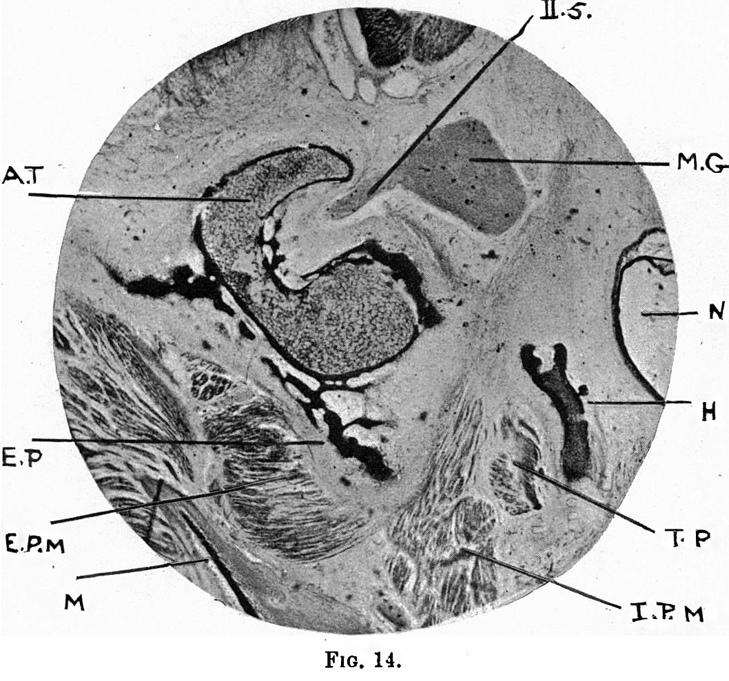
Fig.14, Coronal section of the head of an 80mm embryo
Here the cartilage of the great wing is shown with very characteristic form at this plane of section, appearing somewhat like a half-bent forefinger (A.T.), the concavity being the anterior end of the foramen rotundum; the superior maxillary division of the 5th nerve (I.5) is seen in this concavity running outwards from Meckel's ganglion (M.G.). The cartilage is in a somewhat advanced condition of osification, but its relative size is no greater than in the 30 mm embryo, and its form is quite identical with that in the above-mentioned embryo. Projecting downwards from the lower end of the cartilage, membrane bone of deep black colour is seen forming.the ectochondral external pterygoid plate (E.P.), whilst in the upward direction membrane bone is evident, and it will form, as previously stated, the orbital plate and that part of the great wing which is found in the temporal fossa.
| Historic Disclaimer - information about historic embryology pages |
|---|
 |
|
|
Reference
Fawcett E. Notes on the development of the human sphenoid. (1910) J Anat. Physiol. 44(3): 207-22. PMID 17232842
Cite this page: Hill, M.A. (2024, April 27) Embryology Fawcett 1910 fig14.jpg. Retrieved from https://embryology.med.unsw.edu.au/embryology/index.php/File:Fawcett_1910_fig14.jpg
- © Dr Mark Hill 2024, UNSW Embryology ISBN: 978 0 7334 2609 4 - UNSW CRICOS Provider Code No. 00098G
File history
Click on a date/time to view the file as it appeared at that time.
| Date/Time | Thumbnail | Dimensions | User | Comment | |
|---|---|---|---|---|---|
| current | 08:34, 29 December 2014 | | 1,058 × 989 (271 KB) | Z8600021 (talk | contribs) | {{Fawcett1910_sphenoid_figures}} |
You cannot overwrite this file.
File usage
The following page uses this file: