File:Fawcett1911 fig03.jpg
From Embryology

No higher resolution available.
Fawcett1911_fig03.jpg (600 × 375 pixels, file size: 37 KB, MIME type: image/jpeg)
Fig. 3. Inner view of 19 mm model, Minot embryo
P.P., palatine process; F.P., frontal process; I.O.N., infraorbital nerve; A.D.N., anterior dental nerve; 0.8., orbital surface; O.A.P., buter alveolar process.
| Historic Disclaimer - information about historic embryology pages |
|---|
 |
Reference
Fawcett E. The development of the human maxilla, vomer, and paraseptal cartilages. (1911) J Anat. Physiol. 45(4): 378-405.
Cite this page: Hill, M.A. (2024, April 26) Embryology Fawcett1911 fig03.jpg. Retrieved from https://embryology.med.unsw.edu.au/embryology/index.php/File:Fawcett1911_fig03.jpg
- © Dr Mark Hill 2024, UNSW Embryology ISBN: 978 0 7334 2609 4 - UNSW CRICOS Provider Code No. 00098G
File history
Click on a date/time to view the file as it appeared at that time.
| Date/Time | Thumbnail | Dimensions | User | Comment | |
|---|---|---|---|---|---|
| current | 22:33, 22 May 2017 | | 600 × 375 (37 KB) | Z8600021 (talk | contribs) | |
| 22:32, 22 May 2017 | 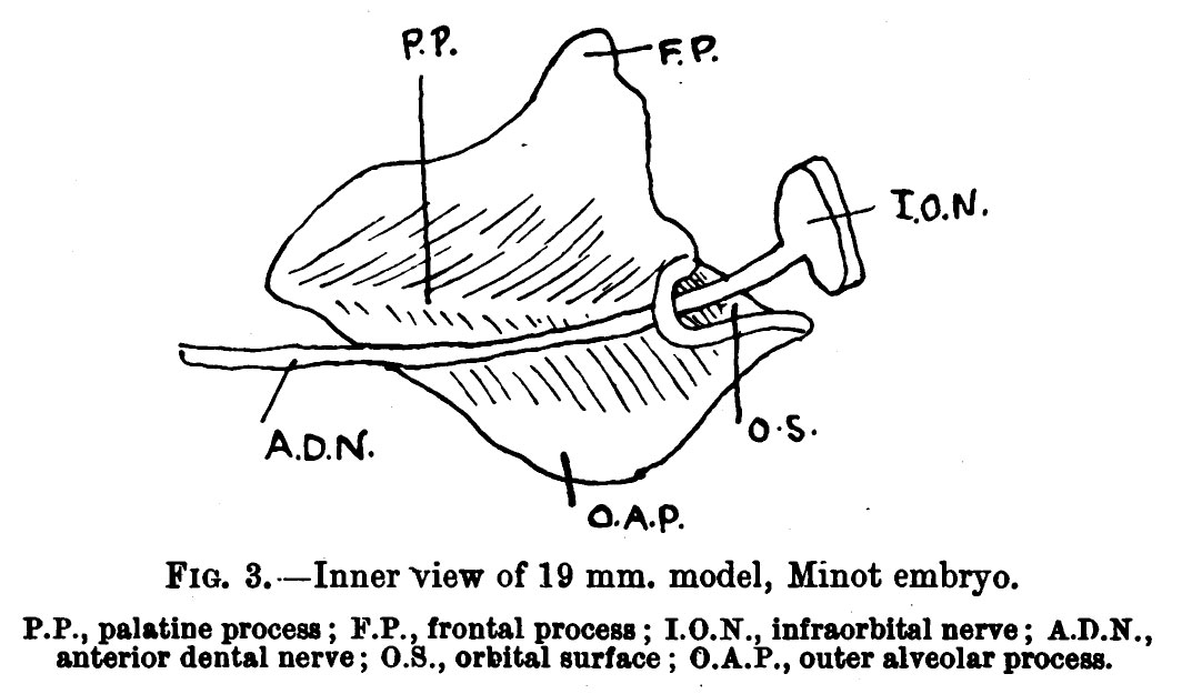 | 1,064 × 625 (95 KB) | Z8600021 (talk | contribs) |
You cannot overwrite this file.
File usage
The following page uses this file: