File:Cullen1916 fig01.jpg

Original file (1,280 × 929 pixels, file size: 354 KB, MIME type: image/jpeg)
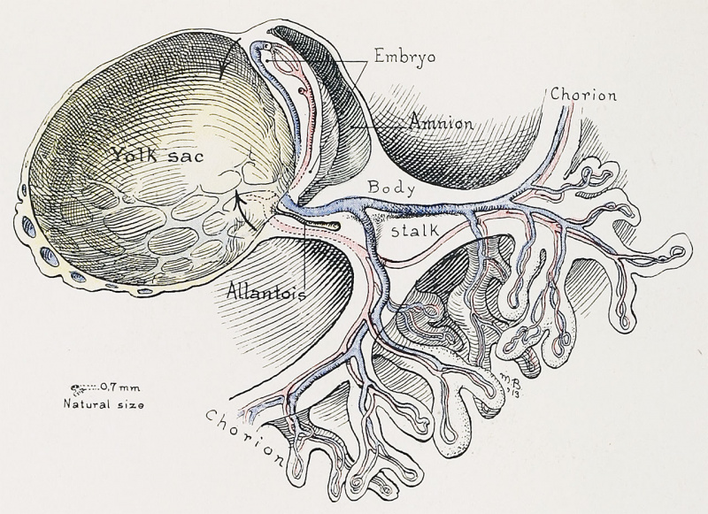
Fig. 1. Sagittal Section Showing a Very Early Stage in the Formation of the Umbilicus and Allantois
(Human embryo, 0.7 mm. long.)
Note the origin of the allantois from the cavity of the yolk-sac. The umbilical region will be formed by a gradua
approximation of the cranial and caudal ends of the yolk-sac, as indicated by the arrows. The embryonic surface of
the yolk-sac will later become the alimentary canal.
| Historic Disclaimer - information about historic embryology pages |
|---|
 |
- Figure Links: 1 Human embryo 0.7 mm | 2 Human embryo 1.7 mm | 3 Human embryo 2.5 mm | 4 Human embryo 3.5 mm | 5 Human embryo 5 mm | 6 Human embryo 7 mm | 7 Human embryo 7 mm | 8 Human embryo 10 mm | 9 Human embryo 12.5 mm | 10 Human embryo 10 mm | 11 Human embryo 23 mm | 12 Human embryo 3 cm | 13 Human embryo 4.5 cm sagittal | 14 Human Embryo 4.5 cm | 15 Human Embryo 5.2 cm | 16 Human Embryo 6.5 cm | 17 Human Embryo 7.5 cm | 18 Human Embryo 9 cm | 19 Human Embryo 10 cm | 20 Human Embryo 12 cm | 21 Human Embryo 12 cm | 22 Human Embryo 12 cm | 23 Human Embryo 12 cm Cord | 28 Fetus Five Months | 30 Ventral Heria | 31 Human Embryo 5.5 cm | 32 Term Human | 33 Term Human | [[Figures
Reference
Cullen TS. Embryology, anatomy, and diseases of the umbilicus together with diseases of the urachus. (1916) W. B. Saunders Company, Philadelphia And London.
Cite this page: Hill, M.A. (2024, April 27) Embryology Cullen1916 fig01.jpg. Retrieved from https://embryology.med.unsw.edu.au/embryology/index.php/File:Cullen1916_fig01.jpg
- © Dr Mark Hill 2024, UNSW Embryology ISBN: 978 0 7334 2609 4 - UNSW CRICOS Provider Code No. 00098G
File history
Click on a date/time to view the file as it appeared at that time.
| Date/Time | Thumbnail | Dimensions | User | Comment | |
|---|---|---|---|---|---|
| current | 15:57, 27 October 2018 | | 1,280 × 929 (354 KB) | Z8600021 (talk | contribs) | |
| 15:56, 27 October 2018 |  | 2,163 × 1,417 (587 KB) | Z8600021 (talk | contribs) | Fig. 1. — Sagittal Section Showing a Very Early Stage in the Formation of the Umbilicus and Allantois. (Human embryo, 0.7 mm. long.) Note the origin of the allantois from the cavity of the yolk-sac. The umbilical region will be formed by a gradua a... |
You cannot overwrite this file.
File usage
The following 3 pages use this file: