File:Control and parthenogenetic canine fetuses.jpg

Original file (660 × 800 pixels, file size: 111 KB, MIME type: image/jpeg)
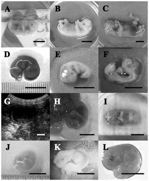
Comparison of control and parthenogenetic canine fetuses
(A–C) The relative size and gross morphology of a control fetus on days 28, 30 and 32 of pregnancy, respectively.
(D–F) Parthenogenetic fetuses on day 28 of pregnancy.
(G) In utero ultrasonograph of parthenogenetic fetuses on day 29 of pregnancy.
(H, I) A parthenogenetic fetus recovered on day 30 of pregnancy. Note that the vascular system is well developed and the external morphology is similar to the control.
(J) Another small parthenogenetic fetus recovered on day 30 of pregnancy.
(K, L) Parthenogenetic fetuses recovered on day 32 of pregnancy.
All scale bars represent 10 mm.
Reference
<pubmed>22905100</pubmed> | PMC3419697 | PLoS One.
Citation: Park JE, Kim MJ, Ha SK, Hong SG, Oh HJ, et al. (2012) Altered Cell Cycle Gene Expression and Apoptosis in Post-Implantation Dog Parthenotes. PLoS ONE 7(8): e41256. doi:10.1371/journal.pone.0041256
Copyright: © 2012 Park et al. This is an open-access article distributed under the terms of the Creative Commons Attribution License, which permits unrestricted use, distribution, and reproduction in any medium, provided the original author and source are credited.
Figure 2. doi:10.1371/journal.pone.0041256.g002
Journal.pone.0041256.g002.jpg
File history
Click on a date/time to view the file as it appeared at that time.
| Date/Time | Thumbnail | Dimensions | User | Comment | |
|---|---|---|---|---|---|
| current | 16:32, 25 September 2012 |  | 660 × 800 (111 KB) | Z8600021 (talk | contribs) | ==Comparison of control and parthenogenetic canine fetuses== (A–C) The relative size and gross morphology of a control fetus on days 28, 30 and 32 of pregnancy, respectively. (D–F) Parthenogenetic fetuses on day 28 of pregnancy. (G) In utero ultr |
You cannot overwrite this file.
File usage
There are no pages that use this file.