File:Conklin 1905 plate05.jpg
Original file (1,521 × 2,000 pixels, file size: 331 KB, MIME type: image/jpeg)
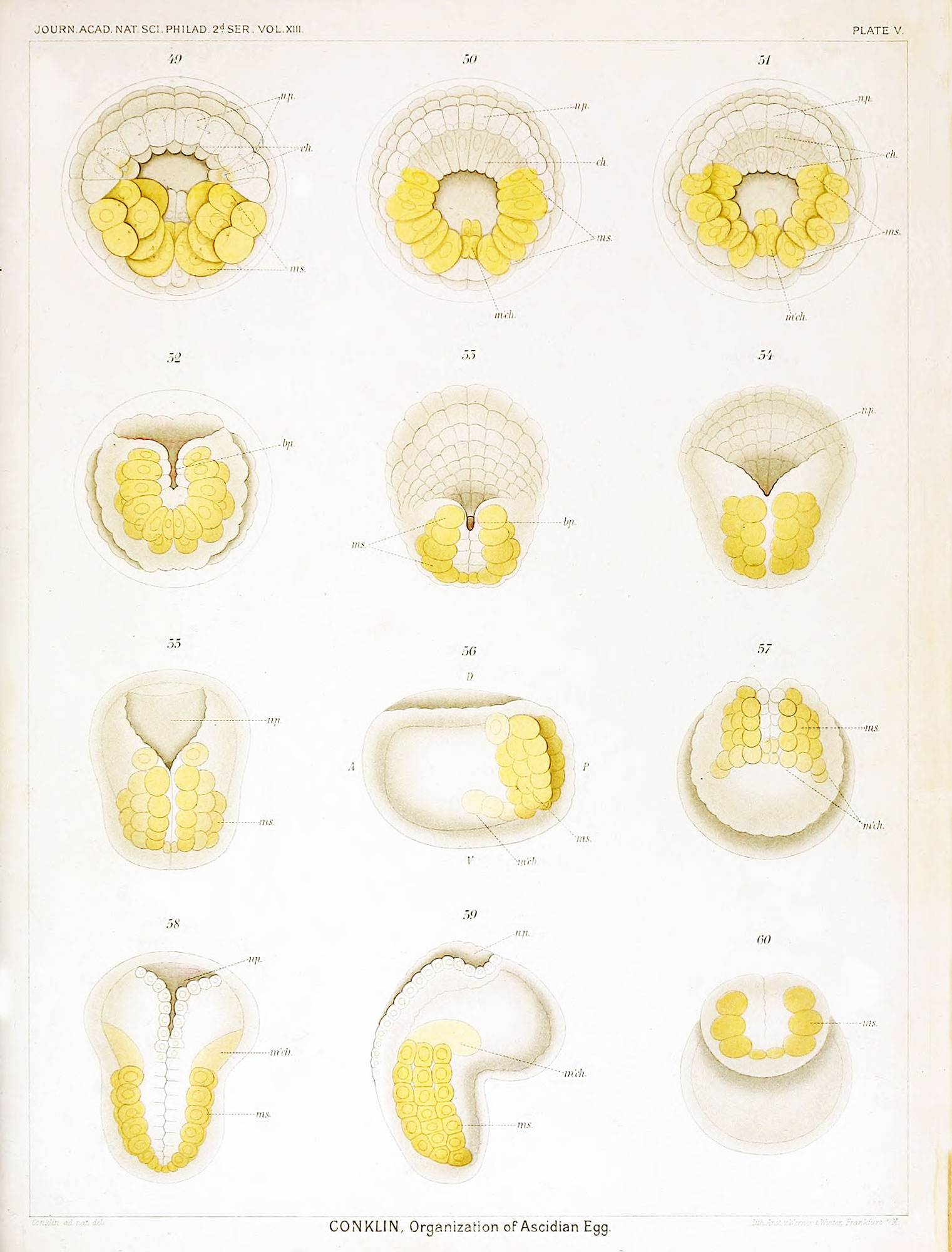
Plate V Living Embryo of Oynthia partita - Gastrula to Tadpole
Fig. 49. Gastrula showing neural plate, chorda and muscle cells; about 176 cells, 96 ectoderm, 32 mesoderm, 20 endoderm, 8 chorda, 8 dorsal neural plate and 12 ventral neural plate cells.
Figs. 50 and 51. Two stages of one embryo, the second drawn about ten minutes after the first; the yellow cells in the mid-line (m'ch.) are mesenchyme cells, the others (ms.) muscle cells.
Fig. 52. Posterior view of elongated gastrula, the blastopore reduced to a narrow slit.
Fig. 53. Dorsal view of similar stage, the blastopore a small opening at the posterior end of a groove
Fig. 54. An older embryo, the blastopore covered by the forward growth of the posterior lips.
Fig. 55. Embryo with inrolling neural plate and with muscle cells arranged in three rows.
Fig. 56. Embryo of about the same stage as that shown in figs. 52 and 53; seen from the left side showing neural groove, three rows of muscle cells which run transversely to the long axis, and a row of mesenchyme cells in the long axis.
Fig. 57. Embryo viewed from posterior end, showing blastopore-raphe with open groove above and with three rows of muscle cells on each side, also a row of mesenchyme cells.
Fig. 58. Young tadpole seen from dorsal side, neural groove open in front and closed behind, smallcelled mesenchyme in front of large muscle cells.
Fig. 59. Same stage as preceding seen from the right side, showing neural groove, mesenchyme and three rows of muscle, cells.
Fig. 60. Tadpole of slightly older stage viewed from the posterior (caudal) end showing on each side three large muscle cells each of which belongs to a row of such cells (cf Fig. 59). These muscle cells are connected across the mid-line at the posterior end by a few small mesenchyme cells.
| Historic Disclaimer - information about historic embryology pages |
|---|
 |
- Conklin Figures: Fig 1-2 | Fig 3-6 | Fig 7-8 | Fig 9-12 | Fig 13-16 | Fig 17-20 | Fig 21-24 | Fig 25-26 | Fig 27-33 | Fig 34-35 | Plate I | Plate II | Plate III | Plate IV | Plate V | Plate VI | Plate VII | Plate VIII | Plate IX | Plate X | Plate XI | Plate XII
Reference
Conklin EG. The Organization and Cell-Lineage of the Ascidian Egg (1905) J. Acad., Nat. Sci. Phila. 13, 1.
Conklin 1905 TOC: I. The Ovarian Egg | II. Maturation and Fertilization | III. Orientation of Egg and Embryo | IV. Cell-Lineage | V. Later Development | VI. Comparisons with A.mphioxus and Amphibia | VII. The Organization of the Egg | Summary | Literature Cited | Explanation of Figures
Cite this page: Hill, M.A. (2024, April 27) Embryology Conklin 1905 plate05.jpg. Retrieved from https://embryology.med.unsw.edu.au/embryology/index.php/File:Conklin_1905_plate05.jpg
- © Dr Mark Hill 2024, UNSW Embryology ISBN: 978 0 7334 2609 4 - UNSW CRICOS Provider Code No. 00098G
File history
Click on a date/time to view the file as it appeared at that time.
| Date/Time | Thumbnail | Dimensions | User | Comment | |
|---|---|---|---|---|---|
| current | 15:47, 19 October 2016 | | 1,521 × 2,000 (331 KB) | Z8600021 (talk | contribs) |
You cannot overwrite this file.
File usage
The following 2 pages use this file: