File:Conklin 1905 fig13-16.jpg
Original file (1,000 × 796 pixels, file size: 144 KB, MIME type: image/jpeg)
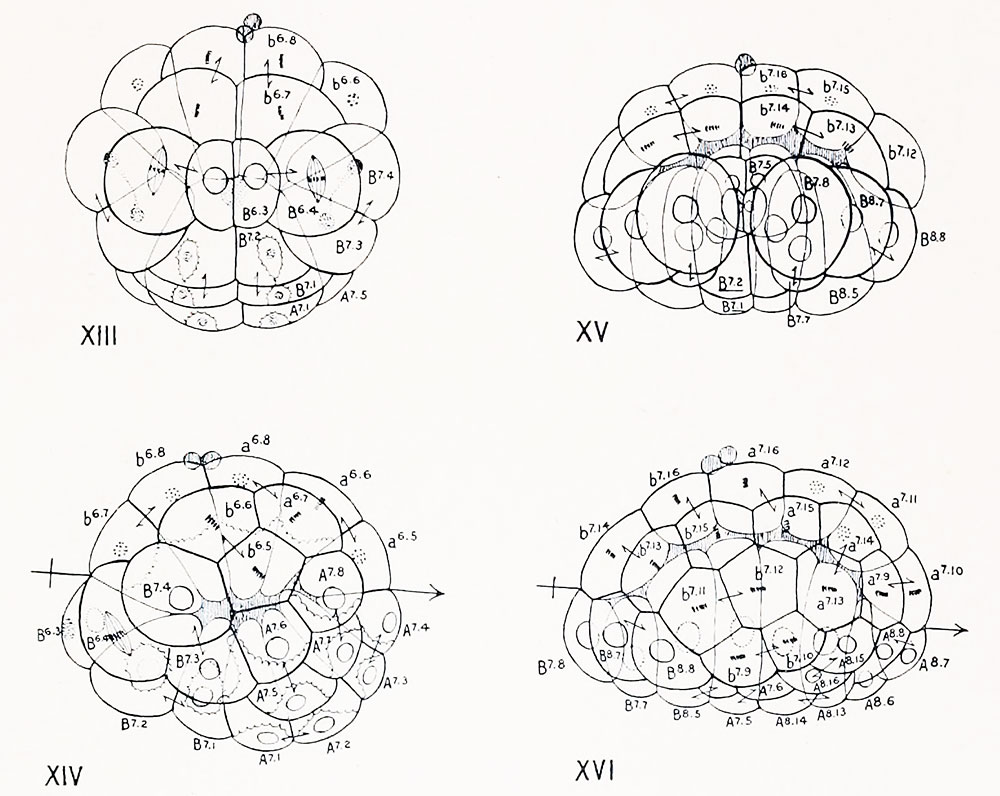
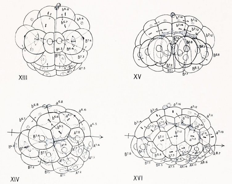
Figs. XIII-XVI Camera drawings of eutire eggs of Ciona intestinalu viewed as transparent objects
Figs. XIII and XV are seen from the posterior pole ; Figs. XIV and XVI from the left side. Figs. XIII and XIV represent a 44-cell stage passing into a 62-cell stage;
Figs. XV and XVI a 76-cell passing into a 110-cell stage. The position of the equator, the boundary between protoplasm and yolk and the segmentation cavity are represented as in the preceding figures.
In Fig. XIV the cells A7-4 and A? 8 are neural plate cells, A?-3 and A" " chorda cells; in Fig. XVI the neural plate cells are A 8 ?, A 8 - 8 , A 8 ."5, A 8 -' 6 , while the chorda cells are A 8 -s, A 8 - 6 , A 8 -'3, A 8 - 1 '!. The crescent cells (mesoderm) are B 6 -3, BM, B?-t, B?-3 (Figs. XIII and XIVl and their dirivatives B7-5, &<>, B7 7, B?- 8 , B 8 -5, B 8 - 6 , BK B 8 - 8 (Fig. XV and XVI). The other cells of the lower hemisphere are endodermal.
In Figs. XIII and XIV the cells bordering the segmentation cavity (seen in optical section) are of about equal height at the two poles; in Figs. XV and XVI the cells at the loVer pole are columnar, those at the upper pole flattened. The polar bodies are present exactly where they appear in the drawings.
| Historic Disclaimer - information about historic embryology pages |
|---|
 |
- Conklin Figures: Fig 1-2 | Fig 3-6 | Fig 7-8 | Fig 9-12 | Fig 13-16 | Fig 17-20 | Fig 21-24 | Fig 25-26 | Fig 27-33 | Fig 34-35 | Plate I | Plate II | Plate III | Plate IV | Plate V | Plate VI | Plate VII | Plate VIII | Plate IX | Plate X | Plate XI | Plate XII
Reference
Conklin EG. The Organization and Cell-Lineage of the Ascidian Egg (1905) J. Acad., Nat. Sci. Phila. 13, 1.
Conklin 1905 TOC: I. The Ovarian Egg | II. Maturation and Fertilization | III. Orientation of Egg and Embryo | IV. Cell-Lineage | V. Later Development | VI. Comparisons with A.mphioxus and Amphibia | VII. The Organization of the Egg | Summary | Literature Cited | Explanation of Figures
Cite this page: Hill, M.A. (2024, April 26) Embryology Conklin 1905 fig13-16.jpg. Retrieved from https://embryology.med.unsw.edu.au/embryology/index.php/File:Conklin_1905_fig13-16.jpg
- © Dr Mark Hill 2024, UNSW Embryology ISBN: 978 0 7334 2609 4 - UNSW CRICOS Provider Code No. 00098G
File history
Click on a date/time to view the file as it appeared at that time.
| Date/Time | Thumbnail | Dimensions | User | Comment | |
|---|---|---|---|---|---|
| current | 15:37, 21 October 2016 | | 1,000 × 796 (144 KB) | Z8600021 (talk | contribs) | |
| 15:36, 21 October 2016 | 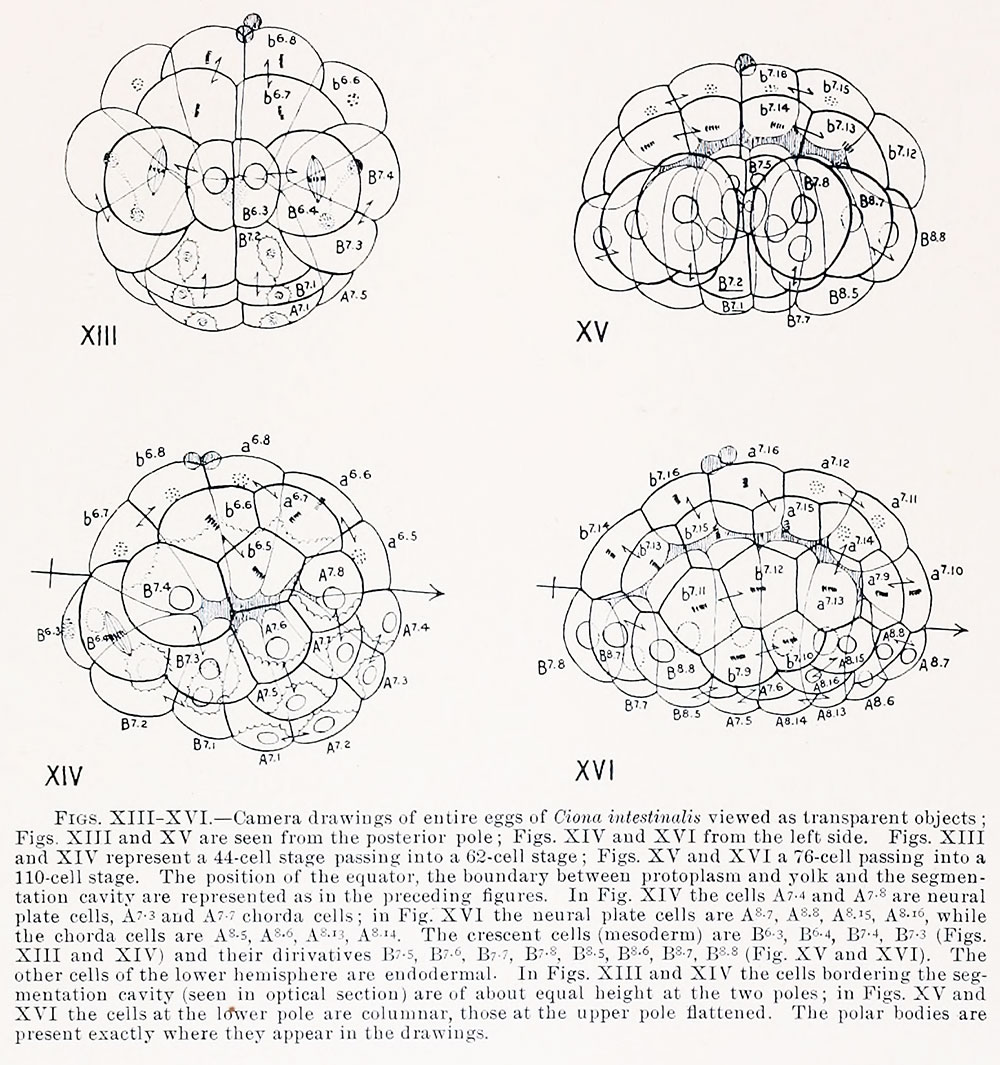 | 1,000 × 1,065 (249 KB) | Z8600021 (talk | contribs) |
You cannot overwrite this file.
File usage
The following 2 pages use this file: