File:Conklin 1905 fig03-06.jpg
Original file (1,000 × 1,007 pixels, file size: 100 KB, MIME type: image/jpeg)
Fig. 5-6
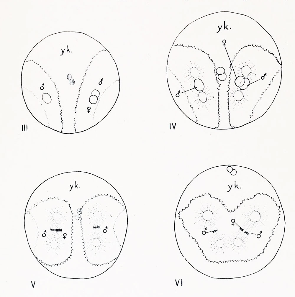
Figs. III-VI. Dispermic eggs of ('. partita; drawn from stained preparations of entire eggs. Figs. III. and V are viewed from the vegetal pole, the polar bodies being seen through the egg. Fig. IV is viewed from the animal pole and Fig. VI from the posterior pole. The boundary between the protoplasm and yolk is indicated by a crenated line; when seen through the egg this boundary is represented by a line of stipples.
| Historic Disclaimer - information about historic embryology pages |
|---|
 |
- Conklin Figures: Fig 1-2 | Fig 3-6 | Fig 7-8 | Fig 9-12 | Fig 13-16 | Fig 17-20 | Fig 21-24 | Fig 25-26 | Fig 27-33 | Fig 34-35 | Plate I | Plate II | Plate III | Plate IV | Plate V | Plate VI | Plate VII | Plate VIII | Plate IX | Plate X | Plate XI | Plate XII
Reference
Conklin EG. The Organization and Cell-Lineage of the Ascidian Egg (1905) J. Acad., Nat. Sci. Phila. 13, 1.
Conklin 1905 TOC: I. The Ovarian Egg | II. Maturation and Fertilization | III. Orientation of Egg and Embryo | IV. Cell-Lineage | V. Later Development | VI. Comparisons with A.mphioxus and Amphibia | VII. The Organization of the Egg | Summary | Literature Cited | Explanation of Figures
Cite this page: Hill, M.A. (2024, April 26) Embryology Conklin 1905 fig03-06.jpg. Retrieved from https://embryology.med.unsw.edu.au/embryology/index.php/File:Conklin_1905_fig03-06.jpg
- © Dr Mark Hill 2024, UNSW Embryology ISBN: 978 0 7334 2609 4 - UNSW CRICOS Provider Code No. 00098G
File history
Click on a date/time to view the file as it appeared at that time.
| Date/Time | Thumbnail | Dimensions | User | Comment | |
|---|---|---|---|---|---|
| current | 17:00, 19 October 2016 | | 1,000 × 1,007 (100 KB) | Z8600021 (talk | contribs) | |
| 17:00, 19 October 2016 | 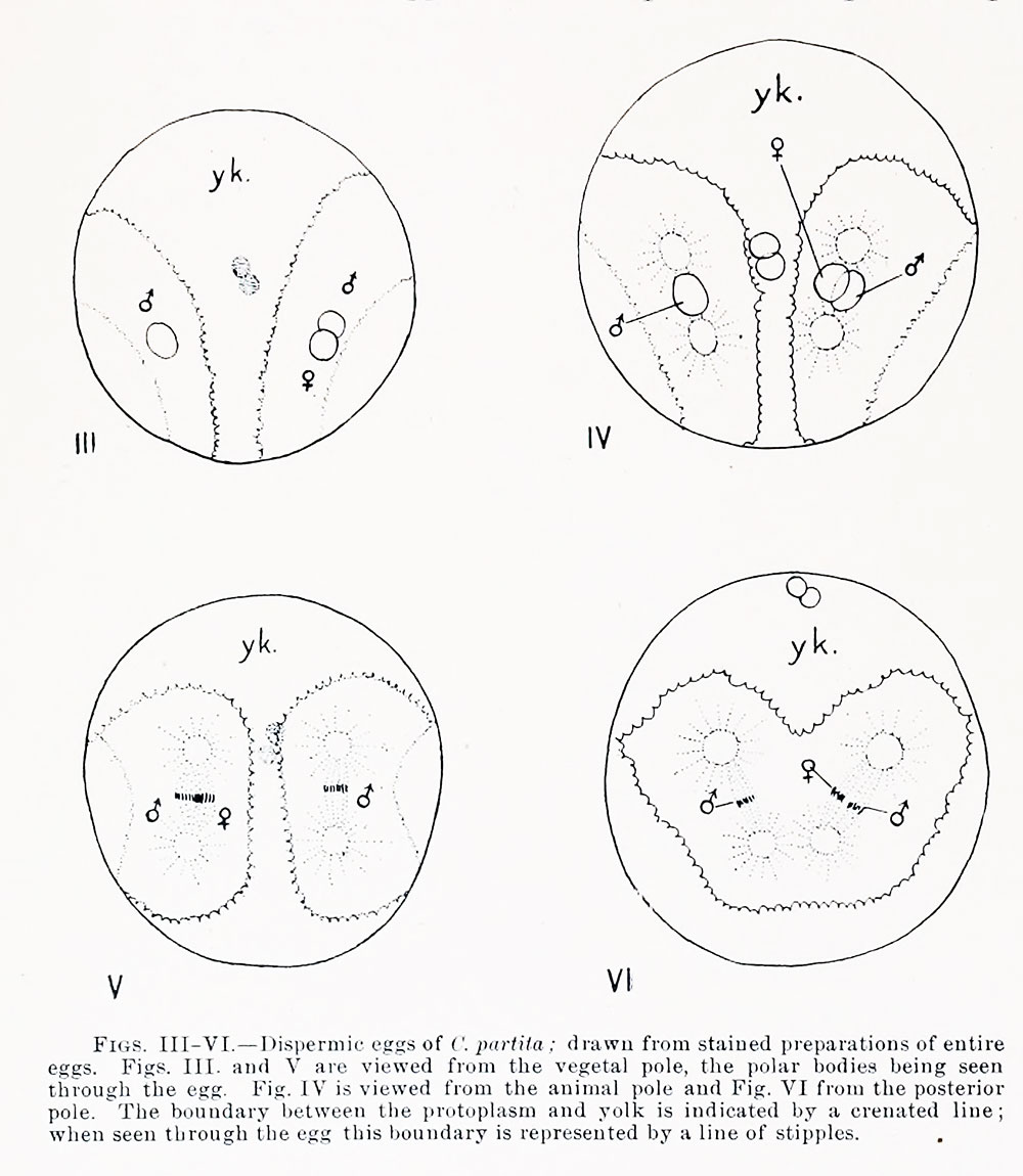 | 1,000 × 1,150 (144 KB) | Z8600021 (talk | contribs) | {{Historic Disclaimer}} {{Conklin1905 figures}} ===Reference=== {{Ref-Conklin1905}} {{Conklin1905 TOC}} {{Footer}} |
You cannot overwrite this file.
File usage
The following 2 pages use this file: