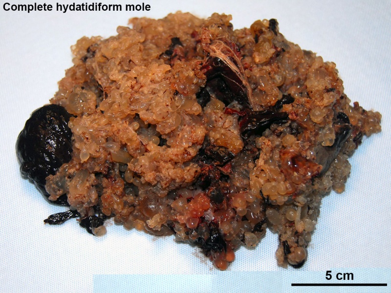
File:Complete hydatidiform mole 05.jpg
From Embryology

Size of this preview: 800 × 600 pixels. Other resolution: 1,280 × 960 pixels.
Original file (1,280 × 960 pixels, file size: 324 KB, MIME type: image/jpeg)
Complete Hydatidiform Mole
O01.0 Complete hydatidiform mole
- Links: hydatidiform mole
Reference
Image: Dr Steven O'Connor (Houston, Texas) - Other embryo images.
Cite this page: Hill, M.A. (2024, April 26) Embryology Complete hydatidiform mole 05.jpg. Retrieved from https://embryology.med.unsw.edu.au/embryology/index.php/File:Complete_hydatidiform_mole_05.jpg
- © Dr Mark Hill 2024, UNSW Embryology ISBN: 978 0 7334 2609 4 - UNSW CRICOS Provider Code No. 00098G
File history
Click on a date/time to view the file as it appeared at that time.
| Date/Time | Thumbnail | Dimensions | User | Comment | |
|---|---|---|---|---|---|
| current | 22:45, 31 May 2016 | | 1,280 × 960 (324 KB) | Z8600021 (talk | contribs) |
You cannot overwrite this file.
File usage
The following page uses this file: