File:Complete hydatidiform mole 03.jpg

Complete_hydatidiform_mole_03.jpg (748 × 560 pixels, file size: 103 KB, MIME type: image/jpeg)
Complete Hydatidiform Mole
The macroscopic image of the expelled mole that has a classical bunch of grapes appearance.
- Hydatidiform Mole Links: Transvaginal ultrasound | Magnetic resonance image | Macroscopic image | Histology | Hydatidiform Mole
Reference
<pubmed>24083041</pubmed>| Case Rep Obstet Gynecol.
Copyright
© 2013 Naoki Matsumoto et al. This is an open access article distributed under the Creative Commons Attribution License, which permits unrestricted use, distribution, and reproduction in any medium, provided the original work is properly cited.
Figure 1 267268.fig.001a.jpg http://www.hindawi.com/journals/criog/2013/267268/fig1/ Image adjusted in size contrast and labelling.
File history
Click on a date/time to view the file as it appeared at that time.
| Date/Time | Thumbnail | Dimensions | User | Comment | |
|---|---|---|---|---|---|
| current | 22:44, 31 May 2016 | | 748 × 560 (103 KB) | Z8600021 (talk | contribs) | Reverted to version as of 05:10, 10 May 2014 |
| 22:44, 31 May 2016 | 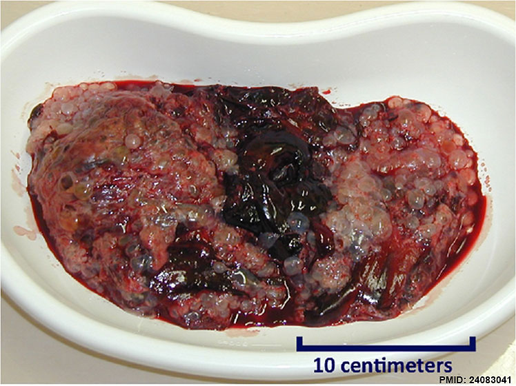 | 748 × 560 (103 KB) | Z8600021 (talk | contribs) | Reverted to version as of 05:10, 10 May 2014 | |
| 22:44, 31 May 2016 | 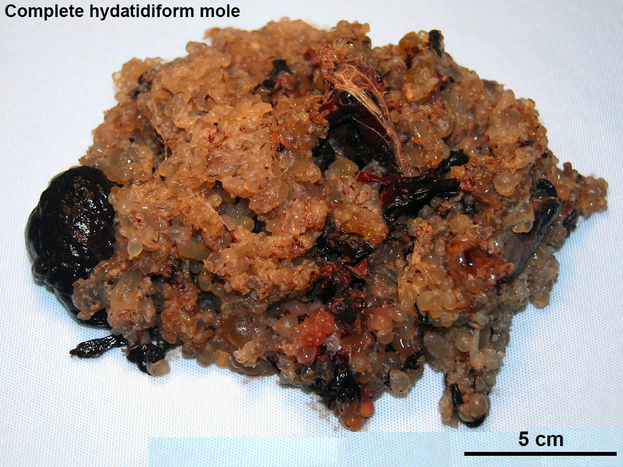 | 1,280 × 960 (324 KB) | Z8600021 (talk | contribs) | ==Complete Hydatidiform Mole== :'''Links:''' Hydatidiform Mole {{SOC}} | |
| 15:10, 10 May 2014 |  | 748 × 560 (103 KB) | Z8600021 (talk | contribs) |
You cannot overwrite this file.
File usage
The following page uses this file: