File:Caudal duplication syndrome.jpg
From Embryology
Caudal_duplication_syndrome.jpg (700 × 599 pixels, file size: 47 KB, MIME type: image/jpeg)
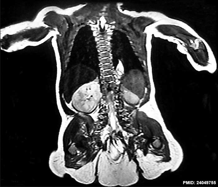
Caudal Duplication Syndrome (MRI)
- Spinal anomalies: Hemi-vertebra at dorsolumbar junction with duplication of vertebral column below that level and sacro-coccygeal agenesis. Spinal dysraphism in the lower lumbar region. Terminal myelocystocele with terminal cord syrinx and evidence of tethered cord with dorsal dermal sinus.
- Duplications of urinary bladder, uterus, and rectum.
- Malrotation of right kidney.
- Enteric duplication cyst in the right iliac fossa.
- Lesion at first and second thoracic vertebra level.
Reference
Sur A, Sardar SK & Paria A. (2013). Caudal duplication syndrome. J Clin Neonatol , 2, 101-2. PMID: 24049755 DOI.
Copyright
File history
Click on a date/time to view the file as it appeared at that time.
| Date/Time | Thumbnail | Dimensions | User | Comment | |
|---|---|---|---|---|---|
| current | 12:04, 24 August 2014 |  | 700 × 599 (47 KB) | Z8600021 (talk | contribs) | ==Caudal duplication syndrome== ===Reference=== <pubmed>24049755</pubmed>| [http://www.jcnonweb.com/article.asp?issn=2249-4847;year=2013;volume=2;issue=2;spage=101;epage=102;aulast=Sur J Clin Neonatol.] ====Copyright==== http://creativecommons.org/l... |
You cannot overwrite this file.
File usage
There are no pages that use this file.
