File:Cardiac muscle EM04.jpg

Original file (1,000 × 680 pixels, file size: 191 KB, MIME type: image/jpeg)
Cardiac Muscle Electron Micrograph
This is a historic (1969) EM showing key features in cardiac muscle ultrastructure. (Stain - Osmium)
Fig 4 Electron micrograph of somewhat more than one sarcomere length of papil- lary muscle fixed at the optimum point on the length-tension curve . The sarcomere length is -2.3y.The thin actin filaments can be seen in the I bands, and both thick and thin fila- ments are discernible in the A band. The prominent M line (M) shows a faint striation with a period of 200 A. The narrow pale regions on either side of the M line demarcate the pseudo H zone (Ps II).At this sarcomere length the tips of the actin filaments are very near the margins of the pseudo H zone. Therefore no true H zone can be seen. X 53,000.
Fig 5 A micrograph of a similar segment from a papillary muscle stretched beyond the optimum length for development of tension . The sarcomere length is noticeably greater (2.6 μ) than in Fig.4, but the length of the A band remains constant . In stretching, the ends of the thin filaments have been pulled away from the M line so that a true H
zone (II) is discernible. X 53,000.
Legend
- Nel - nucleus.
- Mt - Rows of mitochondria appear to divide the contractile substance into myofibril-like units but, unlike the true myofibrils of skeletal muscle, these branch and rejoin and are quite variable in width.
- Mfl - Myofilaments.
- Cap - Capillary.
Original image X 6,700.
Reference
Fawcett DW & McNutt NS. (1969). The ultrastructure of the cat myocardium. I. Ventricular papillary muscle. J. Cell Biol. , 42, 1-45. PMID: 4891913
Copyright
Rockefeller University Press - Copyright Policy This article is distributed under the terms of an Attribution–Noncommercial–Share Alike–No Mirror Sites license for the first six months after the publication date (see http://www.jcb.org/misc/terms.shtml). After six months it is available under a Creative Commons License (Attribution–Noncommercial–Share Alike 4.0 Unported license, as described at https://creativecommons.org/licenses/by-nc-sa/4.0/ ). (More? Help:Copyright Tutorial)
Original article figure (FIG. 4 and 5) has been scaled and rotated.
Cite this page: Hill, M.A. (2024, April 26) Embryology Cardiac muscle EM04.jpg. Retrieved from https://embryology.med.unsw.edu.au/embryology/index.php/File:Cardiac_muscle_EM04.jpg
- © Dr Mark Hill 2024, UNSW Embryology ISBN: 978 0 7334 2609 4 - UNSW CRICOS Provider Code No. 00098G
File history
Click on a date/time to view the file as it appeared at that time.
| Date/Time | Thumbnail | Dimensions | User | Comment | |
|---|---|---|---|---|---|
| current | 23:53, 4 October 2012 | | 1,000 × 680 (191 KB) | Z8600021 (talk | contribs) | |
| 23:52, 4 October 2012 | 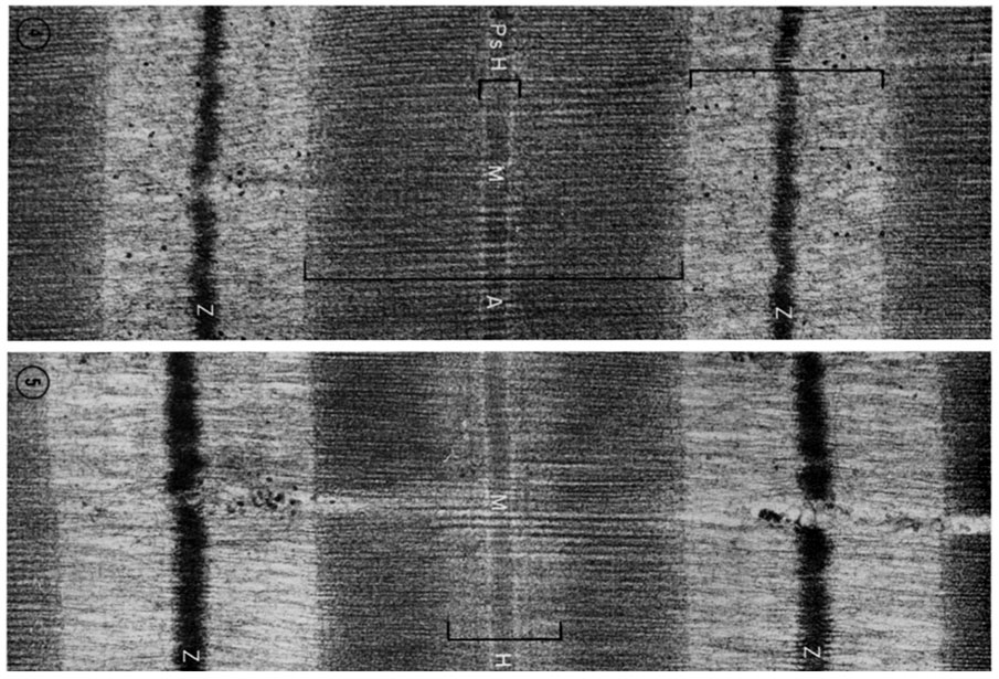 | 905 × 615 (168 KB) | Z8600021 (talk | contribs) | ==Cardiac Muscle Electron Micrograph== This is a historic (1969) EM showing key features in cardiac muscle ultrastructure. Fig 4 Electron micrograph of somewhat more than one sarcomere length of papil- lary muscle fixed at the optimum point on the lengt |
You cannot overwrite this file.
File usage
There are no pages that use this file.